Skip to Content
Merck
All Photos(3)

Key Documents

ABC299

Sigma-Aldrich

Anti-FFAR2 Antibody/GPR43

from rabbit, purified by affinity chromatography

Synonym(s):

Free fatty acid receptor 2, G-protein coupled receptor 43, FFAR2/GPR43

Sign Into View Organizational & Contract Pricing


About This Item

UNSPSC Code:
12352203
eCl@ss:
32160702
NACRES:
NA.41
clone:
polyclonal
application:
IHC
WB
species reactivity:
rat
technique(s):
immunohistochemistry: suitable
western blot: suitable
citations:
13

biological source

rabbit

Quality Level

antibody form

affinity isolated antibody

antibody product type

primary antibodies

clone

polyclonal

purified by

affinity chromatography

species reactivity

rat

species reactivity (predicted by homology)

human (based on 100% sequence homology), mouse

technique(s)

immunohistochemistry: suitable
western blot: suitable

NCBI accession no.

UniProt accession no.

shipped in

wet ice

target post-translational modification

unmodified

Gene Information

human ... FFAR2(2867)

General description

Free fatty acid receptor 2 (FFAR2), also known as G-protein coupled receptor 43 (GPR43), functions as the cell membrane receptor for short chain fatty acids that ultimately stimulates G protein coupled, inhibitory (Gi) signals via the adenylate cyclase pathway. FFAR2/GPR43 is found expressed in leukocytes in the lamina propria of the small intestine mostly but some expression can be also be found in splenocytes, adipose tissue, and enteroendocrine cells of the gut. Interesting, FFAR2/GPR43 because it senses short chain fatty acids which are often key metabolites of gut microbiota, it may serve as a biological sensor to regulate the activity of regulatory T cells and the immune response in diseases such as colitis.

Immunogen

Epitope: Cytoplasmic domain
GST-tagged recombinant protein corresponding to the cytoplasmic domain of human FFAR2/GPR43.

Application

Research Category
Apoptosis & Cancer
Research Sub Category
Apoptosis - Additional
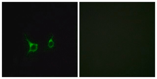
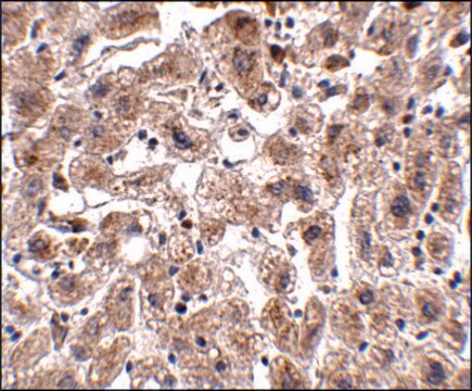
This Anti-FFAR2 Antibody/GPR43 is validated for use in Western Blotting and Immunohistochemistry for the detection of FFAR2/GPR43.
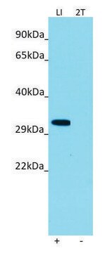
Western Blotting Analysis: 1.0 µg/mL from a representative lot detected FFAR2/GPR43 in 10 µg of RAW264.7 cell lysate.
Immunohistochemistry Analysis: A 1:50 dilution from a representative lot detected FFAR2/GPR43 in mouse kidney and mouse liver tissue.

Quality

Evaluated by Western Blotting in CHEM-1 cell lysate.

Western Blotting Analysis: 1.0 µg/mL of this antibody detected FFAR2/GPR43 in 10 µg of CHEM-1 cell lysate.

Target description

~47 kDa observed. The calculated molecular weight of this protein is 37 kDa, but can be observed at ~47 kDa due to high glycosylation. Uncharacterized band(s) may appear in some lysates.

Physical form

Affinity purified
Purified rabbit polyclonal in buffer containing 0.1 M Tris-Glycine (pH 7.4), 150 mM NaCl with 0.05% sodium azide.

Storage and Stability

Stable for 1 year at 2-8°C from date of receipt.

Other Notes

Concentration: Please refer to lot specific datasheet.

Disclaimer

Unless otherwise stated in our catalog or other company documentation accompanying the product(s), our products are intended for research use only and are not to be used for any other purpose, which includes but is not limited to, unauthorized commercial uses, in vitro diagnostic uses, ex vivo or in vivo therapeutic uses or any type of consumption or application to humans or animals.

Not finding the right product?  

Try our Product Selector Tool.

Storage Class Code

12 - Non Combustible Liquids

WGK

WGK 1

Flash Point(F)

Not applicable

Flash Point(C)

Not applicable


Certificates of Analysis (COA)

Search for Certificates of Analysis (COA) by entering the products Lot/Batch Number. Lot and Batch Numbers can be found on a product’s label following the words ‘Lot’ or ‘Batch’.

Already Own This Product?

Find documentation for the products that you have recently purchased in the Document Library.

Visit the Document Library

Hideaki Fujiwara et al.
Nature communications, 9(1), 3674-3674 (2018-09-12)
Microbiome-derived metabolites influence intestinal homeostasis and regulate graft-versus-host disease (GVHD), but the molecular mechanisms remain unknown. Here we show the metabolite sensor G-protein-coupled receptor 43 (GPR43) is important for attenuation of gastrointestinal GVHD in multiple clinically relevant murine models. GPR43
Zahra Nouri et al.
NPJ biofilms and microbiomes, 8(1), 16-16 (2022-04-06)
Regulating sodium and water balances is crucial for survival of small, desert mammals. Studies demonstrate that the gut microbiota and their metabolites are involved in host energy homeostasis, but little is known on the interactions among salt loading, gut microbiota
Akiko Takakuwa et al.
Nutrients, 11(11) (2019-11-23)
The intestine not only plays a role in fundamental processes in digestion and nutrient absorption, but it also has a role in eliminating ingested pathogenic bacteria and viruses. Paneth cells, which reside at the base of small intestinal crypts, secrete
Ting-Bei Bo et al.
The ISME journal, 13(12), 3037-3053 (2019-08-29)
Gut microbiota play a critical role in orchestrating metabolic homeostasis of the host. However, the crosstalk between host and microbial symbionts in small mammals are rarely illustrated. We used male Brandt's voles (Lasiopodomys brandtii) to test the hypothesis that gut
João R Araújo et al.
Cell host & microbe, 27(3), 358-375 (2020-02-27)
Despite the recognized capacity of the gut microbiota to regulate intestinal lipid metabolism, the role of specific commensal species remains undefined. Here, we aimed to understand the bacterial effectors and molecular mechanisms by which Lactobacillus paracasei and Escherichia coli regulate

Our team of scientists has experience in all areas of research including Life Science, Material Science, Chemical Synthesis, Chromatography, Analytical and many others.

Contact Technical Service