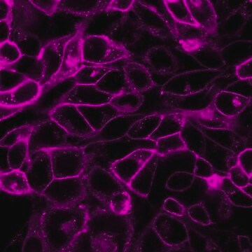
MINI26
PKH26 Red Fluorescent Cell Linker Mini Kit for General Cell Membrane Labeling
Distributed for Phanos Technologies
Synonym(s):
Red PKH membrane label kit
About This Item
Recommended Products
packaging
pkg of 1 kit
manufacturer/tradename
Distributed for Phanos Technologies
storage condition
protect from light
technique(s)
flow cytometry: suitable
fluorescence
λex 551 nm; λem 567 nm (PKH26 dye)
application(s)
cell analysis
detection
detection method
fluorometric
shipped in
ambient
storage temp.
room temp
General description
Application
- labelling microglial, natural killer cells and preadipocytes
- tracking of viable transplanted adipose-derived stem cells
- in vitro proliferation studies of bone marrow stem cells
- general cell membrane labeling, including for in vitro cell labeling and long term in vivo cell tracking.
Linkage
Legal Information
Kit Components Only
- Diluent C 10 mL
- PKH26 Cell Linker in ethanol .1 mL
Signal Word
Danger
Hazard Statements
Precautionary Statements
Hazard Classifications
Eye Irrit. 2 - Flam. Liq. 2
Storage Class Code
3 - Flammable liquids
Flash Point(F)
57.2 °F - closed cup
Flash Point(C)
14 °C - closed cup
Choose from one of the most recent versions:
Already Own This Product?
Find documentation for the products that you have recently purchased in the Document Library.
Customers Also Viewed
Articles
PKH dyes are easy to use and achieve stable, uniform, and reproducible fluorescent labeling of live cells. PKH dyes are non-toxic membrane stains which produce high signal to noise ratio.
Lipophilic cell tracking dyes enable cancer biologists to track tumor and immune cell functions both in vitro and in vivo. Read the article to choose a right membrane dye kit for cell tracking and proliferation monitoring.
Optimal staining is a key component for studying tumorigenesis and progression. Learn useful tips and techniques for dye applications, including examples from recent studies.
A video about how you can use fluorescent cell tracking dyes in combination with flow and image cytometry to study interactions and fates of different cell types in vitro and in vivo.
Our team of scientists has experience in all areas of research including Life Science, Material Science, Chemical Synthesis, Chromatography, Analytical and many others.
Contact Technical Service