Skip to Content
Merck
All Photos(1)

Key Documents

334M-8

Sigma-Aldrich

Cytokeratin (34betaE12) Mouse Monoclonal Antibody

Sign Into View Organizational & Contract Pricing


About This Item

UNSPSC Code:
12352200
NACRES:
NA.41

biological source

mouse

Quality Level

100
500

conjugate

unconjugated

antibody form

culture supernatant

antibody product type

primary antibodies

clone

34betaE12, monoclonal

description

For In Vitro Diagnostic Use in Select Regions (See Chart)

form

buffered aqueous solution

species reactivity

human

packaging

vial of 0.1 mL concentrate (334M-84)
vial of 0.5 mL concentrate (334M-85)
bottle of 1.0 mL predilute (334M-87)
vial of 1.0 mL concentrate (334M-86)
bottle of 7.0 mL predilute (334M-88)

manufacturer/tradename

Cell Marque

technique(s)

immunohistochemistry (formalin-fixed, paraffin-embedded sections): 1:100-1:500

isotype

IgG1κ

control

prostate

shipped in

wet ice

storage temp.

2-8°C

visualization

cytoplasmic

Gene Information

human ... KRT1(3848)

General description

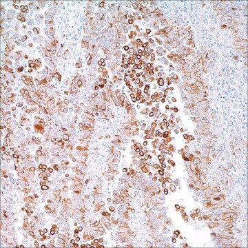
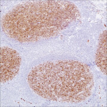
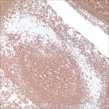
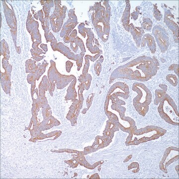
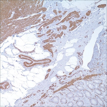
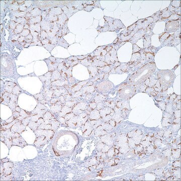
Anti-Cytokeratin, 34betaE12 is an antibody to high molecular weight cytokeratin that reacts with all squamous and ductal epithelium and stains carcinomas. This antibody recognizes cytokeratins 1,5,10, and 14 that are found in complex epithelia. Anti-Cytokeratin, 34betaE12 shows no reactivity with hepatocytes, pancreatic acinar cells, proximal renal tubules, or endometrial glands; there has been no reactivity with cells derived from simple epithelia. Mesenchymal tumors, lymphomas, melanomas, and neural tumors are unreactive with this antibody with some exceptions. Anti-Cytokeratin, 34betaE12 does label myoepithelial cells and has been shown to be useful in distinguishing prostatic adenocarcinoma from hyperplasia of the prostate. This antibody has also been useful in separating benign from malignant intraductal breast proliferations.

Quality


IVD

IVD

IVD

RUO

Linkage

Cytokeratin (34betaE12) Positive Control Slides , Product No. 334S, are available for immunohistochemistry (formalin-fixed, paraffin-embedded sections).

Physical form

Solution in Tris Buffer, pH 7.3-7.7, with 1% BSA and <0.1% Sodium Azide

Preparation Note

Download the IFU specific to your product lot and formatNote: This requires a keycode which can be found on your packaging or product label.

Other Notes

For Technical Service please contact: 800-665-7284 or email: service@cellmarque.com

Legal Information

Cell Marque is a trademark of Merck KGaA, Darmstadt, Germany

Not finding the right product?  

Try our Product Selector Tool.


Certificates of Analysis (COA)

Search for Certificates of Analysis (COA) by entering the products Lot/Batch Number. Lot and Batch Numbers can be found on a product’s label following the words ‘Lot’ or ‘Batch’.

Already Own This Product?

Find documentation for the products that you have recently purchased in the Document Library.

Visit the Document Library

M B Amin et al.
Archives of pathology & laboratory medicine, 118(3), 260-264 (1994-03-01)
Histologic review of 48 radical prostatectomy specimens containing both prostatic adenocarcinoma (PC) and high-grade prostatic intraepithelial neoplasia (PIN) resulted in 23 cases containing neoplastic cribriform gland (CGs) at the periphery or within PC fields. The histologic characteristics of CG PIN
A M Gown et al.
The American journal of pathology, 114(2), 309-321 (1984-02-01)
Monoclonal antibodies generated against different human intermediate filament (IF) proteins were assayed on fixed, embedded tissue by the biotin-avidin-immunoperoxidase method for evaluation of the tissue specificity of these antibodies. An antibody (43 beta E8) made to fibroblast IF protein stains
F P O'Malley et al.
Virchows Archiv. A, Pathological anatomy and histopathology, 417(3), 191-196 (1990-01-01)
There are a number of benign, small-acinar lesions in the prostate gland that may be difficult to differentiate from small-acinar adenocarcinoma. An important diagnostic criterion in this differentiation is the loss of the basal layer in small-acinar adenocarcinoma and its
X J Yang et al.
The American journal of surgical pathology, 23(2), 147-152 (1999-02-16)
Immunohistochemistry with antibodies for high-molecular-weight cytokeratin labels basal cells and is used as an ancillary study in diagnosing prostate carcinoma, which reportedly lacks expression of high-molecular-weight cytokeratin. A recent report questioned the specificity of this marker, describing immunopositivity for high-molecular-weight
K J Wojno et al.
The American journal of surgical pathology, 19(3), 251-260 (1995-03-01)
Basal cell-specific anti-cytokeratin antibody (34 beta E12) decorates the basal cells of benign prostatic epithelium by standard immunohistochemical techniques, whereas adenocarcinoma of the prostate lacks immunoreactivity with this antibody. We reviewed our experience with this antibody to determine its utility

Our team of scientists has experience in all areas of research including Life Science, Material Science, Chemical Synthesis, Chromatography, Analytical and many others.

Contact Technical Service