Saltar al contenido
Merck
Todas las fotos(1)

Key Documents

284M-9

Sigma-Aldrich

Microphthalmia Transcription Factor (MiTF) (C5/D5) Mouse Monoclonal Antibody

Iniciar sesiónpara Ver la Fijación de precios por contrato y de la organización


About This Item

UNSPSC Code:
12352203
NACRES:
NA.41

biological source

mouse

Quality Level

100
500

conjugate

unconjugated

antibody form

culture supernatant

antibody product type

primary antibodies

clone

C5/D5, monoclonal

description

For In Vitro Diagnostic Use in Select Regions (See Chart)

form

buffered aqueous solution

species reactivity

human

packaging

vial of 0.1 mL concentrate (284M-94)
vial of 0.5 mL concentrate (284M-95)
bottle of 1.0 mL predilute (284M-97)
vial of 1.0 mL concentrate (284M-96)
bottle of 7.0 mL predilute (284M-98)

manufacturer/tradename

Cell Marque

technique(s)

immunohistochemistry (formalin-fixed, paraffin-embedded sections): 1:100-1:500

isotype

IgG1

control

melanoma

shipped in

wet ice

storage temp.

2-8°C

visualization

nuclear

Gene Information

human ... MITF(4286)

General description

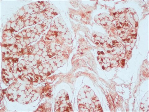
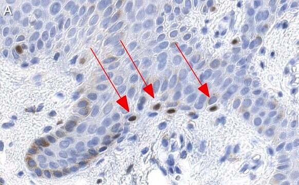
Mi is a transcription factor implicated in pigmentation, bone development and in mast cells. Various forms of Mi exist ranging from 50-70 kD in size. This antibody targets the 52-56 kD range. This antibody has been useful in identifying malignant melanoma, and distinguishing mast cell lesions from lesions of myeloid derivation. A relatively rare class of tumors known as PEComas (tumors showing perivascular epithelioid cell differentiation) express MiTF in a high percentage of cases (~90%).

Quality


IVD

IVD

IVD

RUO

Linkage

Microphthalmia Transcription Factor (MiTF) Positive Control Slides, Product No. 284S, are available for immunohistochemistry (formalin-fixed, paraffin-embedded sections).

Physical form

Solution in Tris Buffer, pH 7.3-7.7, with 1% BSA and <0.1% Sodium Azide

Preparation Note

Download the IFU specific to your product lot and formatNote: This requires a keycode which can be found on your packaging or product label.

Other Notes

For Technical Service please contact: 800-665-7284 or email: service@cellmarque.com

Legal Information

Cell Marque is a trademark of Merck KGaA, Darmstadt, Germany

¿No encuentra el producto adecuado?  

Pruebe nuestro Herramienta de selección de productos.


Certificados de análisis (COA)

Busque Certificados de análisis (COA) introduciendo el número de lote del producto. Los números de lote se encuentran en la etiqueta del producto después de las palabras «Lot» o «Batch»

¿Ya tiene este producto?

Encuentre la documentación para los productos que ha comprado recientemente en la Biblioteca de documentos.

Visite la Librería de documentos

Bernadette Liegl et al.
The American journal of surgical pathology, 32(4), 608-614 (2008-02-19)
PEComas arising in somatic soft tissue or skin are rare. To further characterize the clinicopathologic spectrum, we herein report a series of 10 primary cutaneous PEComas. Eight patients were female and 2 patients were male. The age at presentation ranged
Ilan Weinreb et al.
Virchows Archiv : an international journal of pathology, 450(4), 463-470 (2007-03-23)
Four cases of malignant PEComas were stained with smooth muscle actin, muscle specific actin, desmin, human melanoma black-45, melan-A, microphthalmia transcription factor, S100 and cyclin D1. One case was studied by electron microscopy (EM). Tumour locations were the thigh, elbow
Alberto Righi et al.
International journal of surgical pathology, 16(1), 16-20 (2008-01-22)
We report 2 cases of PEComa, one occurring in the colon of an 11-year-old boy and the other in the bone (fibula) of a 92-year-old woman. Both tumors consisted of nests of large epithelioid cells surrounded by a fibrovascular stroma.
Jason L Hornick et al.
The American journal of surgical pathology, 32(4), 493-501 (2008-01-29)
PEComas (tumors showing perivascular epithelioid cell differentiation) are a family of mesenchymal neoplasms that include angiomyolipoma, clear cell "sugar" tumor of the lung, lymphangiomyomatosis, and a group of uncommon lesions that arise in soft tissue, visceral organs, and skin. We
Steven J Ohsie et al.
Journal of cutaneous pathology, 35(5), 433-444 (2008-04-11)
Melanoma has a wide spectrum of histologic features which mimic epithelial, hematologic, mesenchymal, and neural tumors. Immunohistochemistry has been the primary tool to distinguish melanomas from these other tumors; it has also been studied for use as an adjunct to

Artículos

Immunohistochemistry (IHC) techniques and applications have greatly improved, dermatopathology is still largely based on H&E stained slides.This paper outlines ways in which IHC antibodies can be utilized for dermatopathology.

Nuestro equipo de científicos tiene experiencia en todas las áreas de investigación: Ciencias de la vida, Ciencia de los materiales, Síntesis química, Cromatografía, Analítica y muchas otras.

Póngase en contacto con el Servicio técnico