Saltar al contenido
Merck
Todas las fotos(1)

Key Documents

FCMAB176F

Sigma-Aldrich

Milli-Mark Anti-CD8 -FITC Antibody, clone DK25

clone DK25, Milli-Mark®, from mouse

Iniciar sesiónpara Ver la Fijación de precios por contrato y de la organización


About This Item

UNSPSC Code:
12352203
eCl@ss:
32160702
NACRES:
NA.44

biological source

mouse

Quality Level

conjugate

FITC conjugate

antibody form

purified antibody

antibody product type

primary antibodies

clone

DK25, monoclonal

species reactivity

human

manufacturer/tradename

Milli-Mark®

technique(s)

flow cytometry: suitable

isotype

IgG1κ

NCBI accession no.

UniProt accession no.

shipped in

wet ice

target post-translational modification

unmodified

Gene Information

human ... CD8A(925)

Categorías relacionadas

General description

CD8 is a 68 kDa disulphide-linked, transmembrane glycoprotein. The CD8 molecule serves as a receptor for MHC class I molecules and can mediate a function as a coreceptor in TCR-ligand binding and T-cell activation. Its cytoplasmic part is associated with the p56Ick tyrosine kinase (Nakuchi, 1997). CD8 is expressed by the great majority of cortical thymocytes and approximately 30% of medullary thymocytes, and by class I major histocompatibility complex restricted, mature suppressor/cytotoxic T cells. In addition a proportion of gamma delta T cells and NK cells express CD8 (Nakuchi, 1997). Thus, CD8 is expressed by about 25-35% of peripheral T cells, and about 30% of NK cells express low levels of CD8 (Leong, 1999). The specificity of Anti-CD8, DK25, is equivalent to that of the CD8-clustered antibodies OKT8 and Leu-2a (Stein, 1984).

Specificity

Antibody recognizes Human CD8 antigen.

Application

Milli-Mark Anti-CD8 -FITC Antibody, clone DK25 detects level of CD8 & has been published & validated for use in FC.
Research Category
Inflammation & Immunology
Research Sub Category
Immunoglobulins & Immunology

Quality

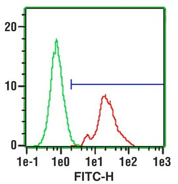
Evaluated by flow cytomatry using human lymphocytes.

Target description

68 kDa (Nakuchi, 1997)

Physical form

Protein A purified
Purified Mouse monoclonal IgG1k conjugated to PE in 0.05M Tris-HCl buffer with 1% BSA, Sodium Azide at 0.097% at pH 7.2

Storage and Stability

Maintain refrigerated at 2-8 °C protected from light in undiluted aliquots for up to 6 months from date of receipt.

Analysis Note

Control
Human Lympnocytes.

Other Notes

Concentration: Please refer to the Certificate of Analysis for the lot-specific concentration.

Legal Information

MILLI-MARK is a registered trademark of Merck KGaA, Darmstadt, Germany

Disclaimer

Unless otherwise stated in our catalog or other company documentation accompanying the product(s), our products are intended for research use only and are not to be used for any other purpose, which includes but is not limited to, unauthorized commercial uses, in vitro diagnostic uses, ex vivo or in vivo therapeutic uses or any type of consumption or application to humans or animals.

¿No encuentra el producto adecuado?  

Pruebe nuestro Herramienta de selección de productos.

Storage Class

12 - Non Combustible Liquids

wgk_germany

WGK 2

flash_point_f

Not applicable

flash_point_c

Not applicable


Certificados de análisis (COA)

Busque Certificados de análisis (COA) introduciendo el número de lote del producto. Los números de lote se encuentran en la etiqueta del producto después de las palabras «Lot» o «Batch»

¿Ya tiene este producto?

Encuentre la documentación para los productos que ha comprado recientemente en la Biblioteca de documentos.

Visite la Librería de documentos

Nicole Marzan-Rivera et al.
iScience, 25(8), 104764-104764 (2022-08-20)
The link between CD4+ T and B cells during immune responses to DENV and ZIKV and their roles in cross-protection during heterologous infection is an active area of research. Here we used CD4+ lymphocyte depletions to dissect the impact of

Nuestro equipo de científicos tiene experiencia en todas las áreas de investigación: Ciencias de la vida, Ciencia de los materiales, Síntesis química, Cromatografía, Analítica y muchas otras.

Póngase en contacto con el Servicio técnico