
추천 제품


색상
clear
응용 분야
hematology
histology
저장 온도
room temp
유사한 제품을 찾으십니까? 방문 제품 비교 안내
일반 설명
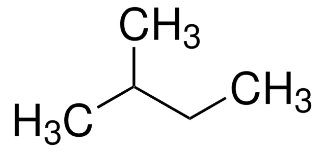
Tissue samples may be snap frozen using PolyFreeze with isopentane and liquid nitrogen, dry ice (slush/slurry or bunker), or in a cryostat.
PolyFreeze media do not cause autofluorescence and can easily be washed away during tissue fixation when rinsed prior to staining. Once rinsed off the slide, there is no trace of the support matrix to interfere with staining or immunohistochemistry (IHC) reactions.
Standard frozen sections at 3–6 μm are easy to obtain and mount on glass slides for drying and/or fixation prior to staining, while thicker sections can be taken as required. Sections will flow freely under an anti-roll device, allowing flat sections to be picked up from the knife-edge. Sections can be either fixed immediately in the fixative of choice or air dried for later fixation and staining. Drying or fixing the slides in the cryostat for specific procedures will not affect the PolyFreeze medium.
um are easy to obtain and mount on glass slides for drying and/or fixation prior to staining, while thicker sections can be taken as required. Sections will flow freely under an anti-roll device, allowing flat sections to be picked up from the knife-edge. Sections can be either fixed immediately in the fixative of choice or air dried for later fixation and staining. Drying or fixing the slides in the cryostat for specific procedures will not affect the PolyFreeze medium.
애플리케이션
A support matrix or form of embedding medium for frozen sectioning. This medium freezes quickly supporting the tissue for sectioning at 3 μ and up with no cracking of the matrix at temperatures from -8 °C to -25 °C. Snap freeze using PolyFreeze with isopentane and liquid nitrogen or dry ice (slush/slurry or bunker). Store specimens frozen in PolyFreeze in liquid nitrogen canisters or in airtight containers in a -80 °C freezer.
Cryo-embedding matrix for frozen specimens.
Cryo-embedding matrix for frozen specimens.
PolyFreeze tissue freezing medium has been used in immunohistochemistry.
PolyFreeze tissue freezing medium has been used in immunohistochemistry.
특징 및 장점
- Colored PolyFreeze (blue, green, yellow) makes differentiating multiple specimens easy during sectioning and staining
- Experience less curling, less ice artifacts, faster freezing with PolyFreeze
저장 및 안정성
Store PolyFreeze tissue freezing medium at room temperature. A cloudy appearance of the unfrozen product has no effect on performance.
Store specimens frozen in PolyFreeze medium in liquid nitrogen canisters or in airtight containers in a -80 0C freezer.
Store specimens frozen in PolyFreeze medium in liquid nitrogen canisters or in airtight containers in a -80 0C freezer.
Storage Class Code
10 - Combustible liquids
WGK
WGK 1
Flash Point (°F)
>230.0 °F
Flash Point (°C)
> 110 °C
이미 열람한 고객

An ex vivo gene therapy approach in X-linked retinoschisis.
Bashar A E, et al.
Molecular Vision, 22, 718-718 (2016)
TGF-β1 and IL-10 expression in epithelial ovarian cancer cell line A2780.
Feng X, et al.
Tropical Journal of Pharmaceutical Research, 14(12), 2179-2185 (2015)
Shweta Kukreja et al.
Developmental biology, 467(1-2), 95-107 (2020-09-14)
The retinotectal system has been extensively studied for investigating the mechanism(s) for topographic map formation. The optic tectum, which is composed of multiple laminae, is the major retino recipient structure in the developing avian brain. Laminar development of the tectum
자사의 과학자팀은 생명 과학, 재료 과학, 화학 합성, 크로마토그래피, 분석 및 기타 많은 영역을 포함한 모든 과학 분야에 경험이 있습니다..
고객지원팀으로 연락바랍니다.