콘텐츠로 건너뛰기
Merck
모든 사진(1)

주요 문서

R129

Sigma-Aldrich

Monoclonal Anti-Ryanodine Receptor antibody produced in mouse

clone 34C, ascites fluid

동의어(들):

Anti-CCO, Anti-KDS, Anti-MHS, Anti-MHS1, Anti-PPP1R137, Anti-RYDR, Anti-RYR, Anti-RYR-1, Anti-SKRR

로그인조직 및 계약 가격 보기


About This Item

MDL number:
UNSPSC 코드:
12352203
NACRES:
NA.41

생물학적 소스

mouse

Quality Level

결합

unconjugated

항체 형태

ascites fluid

항체 생산 유형

primary antibodies

클론

34C, monoclonal

분자량

antigen 565 kDa (non-mammalian vertebrates, doublet at 565 kDa representing the α and β isoforms)

종 반응성

human, rat, rabbit, canine, mink, bovine, frog, fish, primate, chicken, mouse, sheep

기술

direct immunofluorescence: suitable
immunohistochemistry (frozen sections): 1:1,000
immunoprecipitation (IP): suitable
western blot: 1:5,000

동형

IgG1

UniProt 수납 번호

배송 상태

dry ice

저장 온도

−20°C

타겟 번역 후 변형

unmodified

유전자 정보

일반 설명

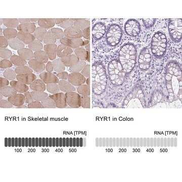
The antibody reacts strongly with RyR-1 (expressed predominantly in skeletal muscle and areas of the brain; also known as the α isoform) and RyR-2 (expressed predominantly in the heart muscle, but also found in stomach, endothelial cells and diffuse areas of the brain; also known as the β isoform). In immunohistochemistry, the antibody stains are consistent with the endoplasmic reticulum localization of the receptor in rat hippocampus.
The ryanodine receptor (RyR) is the channel responsible for the release of Ca2+ from the sarcoplasmic reticulum (SR) in muscle cells and it also plays a role in Ca2+ regulation in non-muscle cells. The RyR exists as a homotetramer and is predicted to have a short cytoplasmic C-terminus and 4-10 transmembrane domains; the remainder of the protein, termed the "foot" region is located in the cytoplasm between the T-tubule and the SR. The mammalian RyR is the product of three different genes: RyR-1, which is expressed predominantly in skeletal muscle and areas of the brain, RyR-2, which is expressed predominantly in the heart muscle but also found in the stomach, endothelial cells and diffuse areas of the brain, and RyR-3 which is found in smooth muscle and the brain (striatum, thalamus and hippocampus).

면역원

partially purified chicken pectoral muscle ryanodine receptors (RyR)

애플리케이션

Applications in which this antibody has been used successfully, and the associated peer-reviewed papers, are given below.
Western Blotting (1 paper)
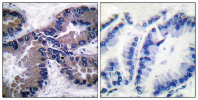
Mouse monoclonal clone 34C anti-Ryanodine receptor is used to tag ryanodine receptors for detection and quantitation by immunocytochemical and immunohistochemical (IHC) techniques. It is used as a probe to determine the presence and roles of ryanodine receptor in studies of calcium flux from the sarcoplasmic reticulum of muscle cells.

물리적 형태

Purified IgG1 antibody containing 0.05% sodium azide.

면책조항

Unless otherwise stated in our catalog or other company documentation accompanying the product(s), our products are intended for research use only and are not to be used for any other purpose, which includes but is not limited to, unauthorized commercial uses, in vitro diagnostic uses, ex vivo or in vivo therapeutic uses or any type of consumption or application to humans or animals.

적합한 제품을 찾을 수 없으신가요?  

당사의 제품 선택기 도구.을(를) 시도해 보세요.

Storage Class Code

10 - Combustible liquids

WGK

nwg

Flash Point (°F)

Not applicable

Flash Point (°C)

Not applicable

개인 보호 장비

Eyeshields, Gloves, multi-purpose combination respirator cartridge (US)


시험 성적서(COA)

제품의 로트/배치 번호를 입력하여 시험 성적서(COA)을 검색하십시오. 로트 및 배치 번호는 제품 라벨에 있는 ‘로트’ 또는 ‘배치’라는 용어 뒤에서 찾을 수 있습니다.

이 제품을 이미 가지고 계십니까?

문서 라이브러리에서 최근에 구매한 제품에 대한 문서를 찾아보세요.

문서 라이브러리 방문

J A Airey et al.
The Journal of biological chemistry, 265(24), 14187-14194 (1990-08-25)
We report evidence for two foot protein isoforms in chicken pectoral muscle. (i) Two polypeptides with molecular masses of approximately 500 kDa copurify with [3H]ryanodine binding. (ii) Both polypeptides are associated with oligomeric proteins similar in size to the mammalian
Janet E Jeffrey et al.
Arthritis research & therapy, 9(6), R129-R129 (2007-12-22)
This study investigated the release of prostaglandin E2 (PGE2) from cartilage following an impact load in vitro and the possible chondroprotective effect of cyclooxygenase-2 (COX-2) inhibition using non-steroidal anti-inflammatory drugs (NSAIDs). Explants of human articular cartilage were subjected to a
Morten Hostrup et al.
Journal of applied physiology (Bethesda, Md. : 1985), 119(5), 475-486 (2015-07-15)
The study was a randomized placebo-controlled trial investigating mechanisms by which chronic β2-adrenergic stimulation enhances muscle force and power output during maximal cycle ergometer exercise in young men. Eighteen trained men were assigned to an experimental group [oral terbutaline 5
Michele Scorzeto et al.
PloS one, 8(10), e74919-e74919 (2013-10-08)
Mitochondrial calcium handling and its relation with calcium released from sarcoplasmic reticulum (SR) in muscle tissue are subject of lively debate. In this study we aimed to clarify how the SR determines mitochondrial calcium handling using dCASQ-null mice which lack
Vanessa Schartner et al.
Acta neuropathologica, 133(4), 517-533 (2016-12-25)
Muscle contraction upon nerve stimulation relies on excitation-contraction coupling (ECC) to promote the rapid and generalized release of calcium within myofibers. In skeletal muscle, ECC is performed by the direct coupling of a voltage-gated L-type Ca2+ channel (dihydropyridine receptor; DHPR)

자사의 과학자팀은 생명 과학, 재료 과학, 화학 합성, 크로마토그래피, 분석 및 기타 많은 영역을 포함한 모든 과학 분야에 경험이 있습니다..

고객지원팀으로 연락바랍니다.