MBD0029
Porphyromonas gingivalis FISH probe - Cy3
Probe for fluorescence in situ hybridization (FISH),20 μM in water
동의어(들):
In-situ hybridization, bacterial fish probes, fish analysis, fish probes, fish technology, fish testing, fluorescence in situ hybridization, fluorescent in situ hybridization, in situ hybridization
로그인조직 및 계약 가격 보기
모든 사진(3)
About This Item
UNSPSC 코드:
12352200
NACRES:
NA.55
추천 제품
일반 설명
Fluorescent In Situ Hybridization technique (FISH) is based on the hybridization of fluorescent labeled oligonucleotide probe to a specific complementary DNA or RNA
sequence in whole and intact cells.1 Microbial FISH allows the visualization, identification and isolation of bacteria due to recognition of ribosomal RNA also in unculturable samples.2
FISH technique can serve as a powerful tool in the microbiome research field by allowing the observation of native microbial populations in diverse microbiome environments, such as samples from human origin (blood3 and tissue4), microbial ecology (solid biofilms5 and aquatic systems6) and plants7. It is strongly recommended to include positive and negative controls in FISH assays to ensure specific binding of the probe of interest and appropriate protocol conditions. We offer positive (MBD0032/33) and negative control (MBD0034/35) probes, that accompany the specific probe of interest.
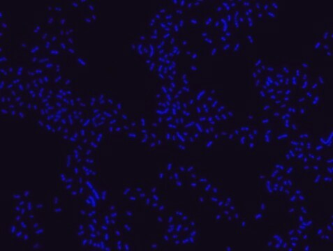
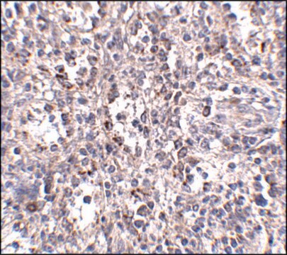
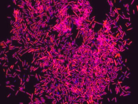
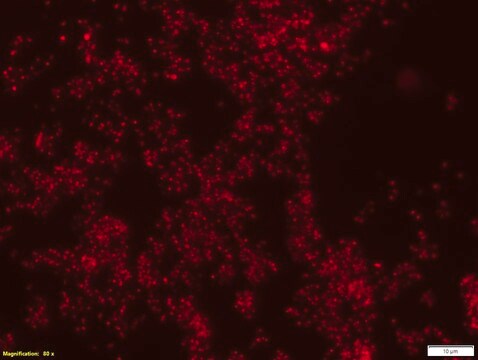
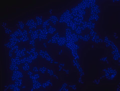
Porphyromonas gingivalis probe specifically recognizes P. gingivalis cells. P. gingivalis, is a gram negative bacterium which is an etiologic agent of adult periodontitis, a chronic inflammatory disease characterized by the destruction of the supportive tissue surrounding teeth. Studies have shown that LPS from P. gingivalis plays an important role in this disease.8-11 The association of the oral microbiota, including P. gingivalis, with various pathological states has been reported. These include development of Alzheimer′s disease12, role in oral cancers13, preterm birth14 and rheumatoid arthritis15. FISH technique was successfully used to identify P. gingivalis with the probe in various samples such as pure culture (as described in the figure legends), dental implants16,17 , periapical tooth lesions18, saliva19, brain tissue20, gingival and aortic tissues21, biofilms from voice prostheses22, subgingival biofilm23, aortic wall tissue24, and infected HeLa cells25. Moreover, FISH can be implicated to identify P. gingivalis in tumor tissue26, multispecies biofilm27, multispecies oral biofilms28 and pure culture and buccal epithelial cells29.
애플리케이션
Probe for fluorescence in situ hybridization (FISH),recognizes Porphyromonas gingivalis cells
특징 및 장점
- Visualize, identify and isolate Porphyromonas gingivalis cells.
- Observe native P. gingivalis cell populations in diverse microbiome environments.
- Specific, sensitive and robust identification of P. gingivalis in bacterial mixed population.
- Specific, sensitive and robust identification even when P. gingivalis is in low abundance in the sample.
- FISH can complete PCR based detection methods by avoiding contaminant bacteria detection.
- Provides information on P. gingivalis morphology and allows to study biofilm architecture.
- Identify P. gingivalis in clinical samples such as, tumor and brain tissues (for example in formalin-fixed paraffin-embedded (FFPE) samples), saliva and oral cavity and medical equipment such as, dental implants and voice prostheses.
- The ability to detect P. gingivalis in its natural habitat is an essential tool for studying host-microbiome interaction.
Storage Class Code
12 - Non Combustible Liquids
WGK
nwg
Flash Point (°F)
Not applicable
Flash Point (°C)
Not applicable
시험 성적서(COA)
제품의 로트/배치 번호를 입력하여 시험 성적서(COA)을 검색하십시오. 로트 및 배치 번호는 제품 라벨에 있는 ‘로트’ 또는 ‘배치’라는 용어 뒤에서 찾을 수 있습니다.
Érika Gomes Sarmento et al.
Food research international (Ottawa, Ont.), 116, 1282-1288 (2019-02-06)
Probiotics are widely used in the food industry and may affect the oral microbiota. This study aimed to evaluate the effect of petit-suisse plus probiotic on the microbiota of children's saliva. Strawberry flavor petit-suisse cheese plus green banana flour without
Nadine Kommerein et al.
PloS one, 13(5), e0196967-e0196967 (2018-05-18)
Peri-implant infections are the most common cause of implant failure in modern dental implantology. These are caused by the formation of biofilms on the implant surface and consist of oral commensal and pathogenic bacteria, which harm adjacent soft and hard
Pia T Sunde et al.
Microbiology (Reading, England), 149(Pt 5), 1095-1102 (2003-05-02)
Whether micro-organisms can live in periapical endodontic lesions of asymptomatic teeth is under debate. The aim of the present study was to visualize and identify micro-organisms within periapical lesions directly, using fluorescence in situ hybridization (FISH) in combination with epifluorescence
Virulence factors of Porphyromonas gingivalis.
S C Holt et al.
Periodontology 2000, 20, 168-238 (1999-10-16)
Alice Harding et al.
Frontiers in aging neuroscience, 9, 398-398 (2017-12-19)
Longitudinal monitoring of patients suggests a causal link between chronic periodontitis and the development of Alzheimer's disease (AD). However, the explanation of how periodontitis can lead to dementia remains unclear. A working hypothesis links extrinsic inflammation as a secondary cause
자사의 과학자팀은 생명 과학, 재료 과학, 화학 합성, 크로마토그래피, 분석 및 기타 많은 영역을 포함한 모든 과학 분야에 경험이 있습니다..
고객지원팀으로 연락바랍니다.
