추천 제품
생물학적 소스
mouse
Quality Level
결합
unconjugated
항체 형태
ascites fluid
항체 생산 유형
primary antibodies
클론
AA6, monoclonal
포함
15 mM sodium azide
종 반응성
human, feline, bovine, hamster, chicken, mouse, rat
기술
microarray: suitable
western blot: 1:500 using a fresh total rat brain extract or an enriched microtubule protein preparation
동형
IgG1
UniProt 수납 번호
배송 상태
dry ice
저장 온도
−20°C
타겟 번역 후 변형
unmodified
유전자 정보
human ... MAP1B(4131)
mouse ... Mtap1b(17755)
rat ... Map1b(29456)
일반 설명
MAP1b is a microtubule associated protein that interacts with actin filaments. MAP1b regulates microtubule stabilization and the development of axons. It may also act as a scaffolding protein Addition of antibody to microtubule proteins before polymerization in immunoassays results in abnormally short (but otherwise morphologically normal) microtubules. Immunohistochemical staining of brain tissue with the product shows selective labeling of dendritic trees throughout the brain. Monoclonal Anti-MAP1b does not react with tubulin of other microtubule associated proteins.
Monoclonal Anti-MAP1b (MAP5) (mouse IgG1 isotype) is derived from the hybridoma produced by the fusion of mouse myeloma cells and splenocytes from an immunized mouse. Microtubule-associated protein (MAP1B) is the major microtubule associated protein in developing brain which changes its expression during development. In the new born rat brain, it is a major component of microtubules but in the adult its level is ten-fold lower.
특이성
Monoclonal Anti-MAP1b antibody is specific for MAP1b in humans, mice, rats, bovines, cats, hamsters and chickens. The antibody does not cross-react with other MAPs or tubulin. Addition of the antibody to microtubule proteins before polymerization results in abnormally short (but otherwise morphologically normal) microtubules. In immunohistochemical staining of brain tissue, the antibody shows selective labeling of neurons with stronger staining of axons, dendrites and cell bodies.
면역원
rat brain microtubule-associated proteins (MAPs)
애플리케이션
Monoclonal Anti-MAP1b antibody produced in mouse has been used in:
- immunohistochemistry
- immunostaining
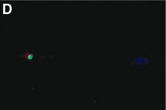
- fluorescence microscopy
생화학적/생리학적 작용
Microtubule-associated protein (MAP1B) is the first MAP to appear in growing axons during development as it is present from the first emergence of the nascent axon from the cell body. Monoclonal Anti-MAP1b may be used to study MAP expression and cytological localization both in tissues and cell lines, under different developmental and environmental circumstances.
면책조항
Unless otherwise stated in our catalog or other company documentation accompanying the product(s), our products are intended for research use only and are not to be used for any other purpose, which includes but is not limited to, unauthorized commercial uses, in vitro diagnostic uses, ex vivo or in vivo therapeutic uses or any type of consumption or application to humans or animals.
적합한 제품을 찾을 수 없으신가요?
당사의 제품 선택기 도구.을(를) 시도해 보세요.
Storage Class Code
10 - Combustible liquids
WGK
WGK 2
Flash Point (°F)
Not applicable
Flash Point (°C)
Not applicable
시험 성적서(COA)
제품의 로트/배치 번호를 입력하여 시험 성적서(COA)을 검색하십시오. 로트 및 배치 번호는 제품 라벨에 있는 ‘로트’ 또는 ‘배치’라는 용어 뒤에서 찾을 수 있습니다.
A Matus et al.
Journal of neurochemistry, 49(3), 714-720 (1987-09-01)
The influence on microtubule assembly in vitro of monoclonal antibodies against microtubule-associated proteins (MAPs) was studied. Light scattering was used for measuring net polymer formation and electron microscopy for determining the influence of antibodies on microtubule morphology. Control experiments showed
Human mesenchymal precursor cells (Stro-1+) from spinal cord injury patients improve functional recovery and tissue sparing in an acute spinal cord injury rat model
Hodgetts SI, et al.
Cell Transplantation, 22(3), 393-412 (2013)
Association of microtubule-associated protein (MAP1B) with growing axons in cultured hippocampal neurons
Fischer I and Romano CG
Molecular and Cellular Neurosciences, 2(1), 39-51 (1991)
Rebecca L Anderson et al.
The Journal of comparative neurology, 447(3), 218-233 (2002-05-02)
Visceromotor neurons in mammalian prevertebral sympathetic ganglia receive convergent synaptic inputs from spinal preganglionic neurons and peripheral intestinofugal neurons projecting from the enteric plexuses. Vasomotor neurons in the same ganglia receive only preganglionic inputs. How this pathway-specific pattern of connectivity
J B Denny
Journal of neurocytology, 20(8), 627-636 (1991-08-01)
A monoclonal antibody was used to determine both the expression of the microtubule-associated protein MAP5 in cultured foetal rat hippocampal neurons as a function of culture age and the cellular distribution of the protein. When cultures at days 2 and
자사의 과학자팀은 생명 과학, 재료 과학, 화학 합성, 크로마토그래피, 분석 및 기타 많은 영역을 포함한 모든 과학 분야에 경험이 있습니다..
고객지원팀으로 연락바랍니다.