
추천 제품


생물학적 소스
rabbit
Quality Level
결합
unconjugated
항체 형태
affinity isolated antibody
항체 생산 유형
primary antibodies
클론
polyclonal
제품 라인
Prestige Antibodies® Powered by Atlas Antibodies
양식
buffered aqueous glycerol solution
종 반응성
human
향상된 검증
independent
orthogonal RNAseq
Learn more about Antibody Enhanced Validation
기술
immunoblotting: 0.04-0.4 μg/mL
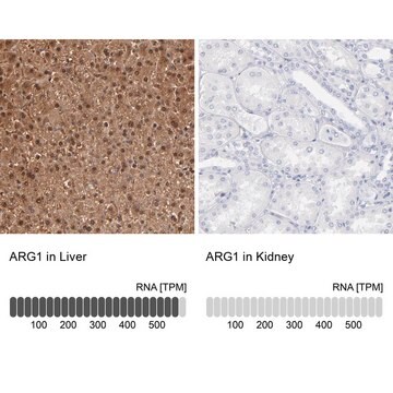
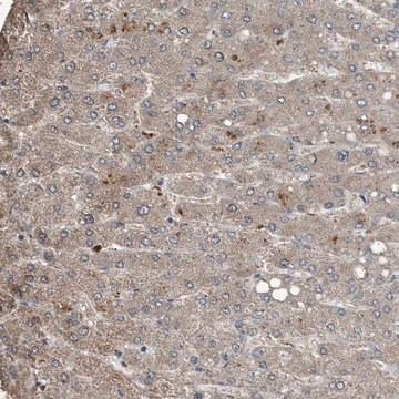
immunohistochemistry: 1:2500-1:5000
면역원 서열
GLRDVDPGEHYILKTLGIKYFSMTEVDRLGIGKVMEETLSYLLGRKKRPIHLSFDVDGLDPSFTPATGTPVVGGLTYREGLYITEEIYKTGLLSGLDIMEVNPSLGKTPEEVTRTVNTAVAITL
UniProt 수납 번호
배송 상태
wet ice
저장 온도
−20°C
타겟 번역 후 변형
unmodified
유전자 정보
human ... ARG1(383)
면역원
Arginase-1 recombinant protein epitope signature tag (PrEST)
애플리케이션
All Prestige Antibodies Powered by Atlas Antibodies are developed and validated by the Human Protein Atlas (HPA) project and as a result, are supported by the most extensive characterization in the industry.
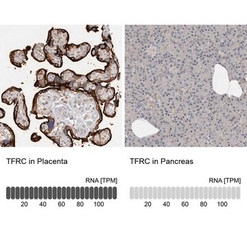
The Human Protein Atlas project can be subdivided into three efforts: Human Tissue Atlas, Cancer Atlas, and Human Cell Atlas. The antibodies that have been generated in support of the Tissue and Cancer Atlas projects have been tested by immunohistochemistry against hundreds of normal and disease tissues and through the recent efforts of the Human Cell Atlas project, many have been characterized by immunofluorescence to map the human proteome not only at the tissue level but now at the subcellular level. These images and the collection of this vast data set can be viewed on the Human Protein Atlas (HPA) site by clicking on the Image Gallery link. We also provide Prestige Antibodies® protocols and other useful information.
The Human Protein Atlas project can be subdivided into three efforts: Human Tissue Atlas, Cancer Atlas, and Human Cell Atlas. The antibodies that have been generated in support of the Tissue and Cancer Atlas projects have been tested by immunohistochemistry against hundreds of normal and disease tissues and through the recent efforts of the Human Cell Atlas project, many have been characterized by immunofluorescence to map the human proteome not only at the tissue level but now at the subcellular level. These images and the collection of this vast data set can be viewed on the Human Protein Atlas (HPA) site by clicking on the Image Gallery link. We also provide Prestige Antibodies® protocols and other useful information.
특징 및 장점
Prestige Antibodies® are highly characterized and extensively validated antibodies with the added benefit of all available characterization data for each target being accessible via the Human Protein Atlas portal linked just below the product name at the top of this page. The uniqueness and low cross-reactivity of the Prestige Antibodies® to other proteins are due to a thorough selection of antigen regions, affinity purification, and stringent selection. Prestige antigen controls are available for every corresponding Prestige Antibody and can be found in the linkage section.
Every Prestige Antibody is tested in the following ways:
Every Prestige Antibody is tested in the following ways:
- IHC tissue array of 44 normal human tissues and 20 of the most common cancer type tissues.
- Protein array of 364 human recombinant protein fragments.
결합
Corresponding Antigen APREST83045
물리적 형태
Solution in phosphate-buffered saline, pH 7.2, containing 40% glycerol and 0.02% sodium azide
법적 정보
Prestige Antibodies is a registered trademark of Merck KGaA, Darmstadt, Germany
면책조항
Unless otherwise stated in our catalog or other company documentation accompanying the product(s), our products are intended for research use only and are not to be used for any other purpose, which includes but is not limited to, unauthorized commercial uses, in vitro diagnostic uses, ex vivo or in vivo therapeutic uses or any type of consumption or application to humans or animals.
적합한 제품을 찾을 수 없으신가요?
당사의 제품 선택기 도구.을(를) 시도해 보세요.
Storage Class Code
10 - Combustible liquids
WGK
WGK 1
Flash Point (°F)
Not applicable
Flash Point (°C)
Not applicable
가장 최신 버전 중 하나를 선택하세요:
Jana de Boniface et al.
Oncoimmunology, 1(8), 1305-1312 (2012-12-18)
Arginase 1 (ARG1) is an important enzyme in amino acid metabolism that also exerts immunoregulatory function. High ARG1 expression, which is associated with cell cycle arrest and functional unresponsiveness in T cells, has been observed after trauma, infections and in
Yichen Xu et al.
Nature medicine, 25(2), 301-311 (2019-01-16)
Cancer cells develop mechanisms to escape immunosurveillance, among which modulating the expression of immune suppressive messenger RNAs is most well-documented. However, how this is molecularly achieved remains largely unresolved. Here, we develop an in vivo mouse model of liver cancer
Xiangdong Wang et al.
Cancer & metabolism, 11(1), 1-1 (2023-01-14)
Arginase-1 (ARG1), a urea cycle-related enzyme, catalyzes the hydrolysis of arginine to urea and ornithine, which regulates the proliferation, differentiation, and function of various cells. However, it is unclear whether ARG1 controls the progression and malignant alterations of colon cancer.
Takahiro Hashimoto-Kataoka et al.
Proceedings of the National Academy of Sciences of the United States of America, 112(20), E2677-E2686 (2015-05-06)
IL-6 is a multifunctional proinflammatory cytokine that is elevated in the serum of patients with pulmonary arterial hypertension (PAH) and can predict the survival of patients with idiopathic PAH (IPAH). Previous animal experiments and clinical human studies indicate that IL-6
Tamar Geiger et al.
Molecular & cellular proteomics : MCP, 12(6), 1709-1722 (2013-02-26)
Identifying the building blocks of mammalian tissues is a precondition for understanding their function. In particular, global and quantitative analysis of the proteome of mammalian tissues would point to tissue-specific mechanisms and place the function of each protein in a
Global Trade Item Number
| SKU | GTIN |
|---|---|
| HPA024006-100UL | 4061837126932 |
| HPA024006-25UL | 4061841413905 |
자사의 과학자팀은 생명 과학, 재료 과학, 화학 합성, 크로마토그래피, 분석 및 기타 많은 영역을 포함한 모든 과학 분야에 경험이 있습니다..
고객지원팀으로 연락바랍니다.