
추천 제품


생물학적 소스
rabbit
결합
unconjugated
항체 형태
affinity isolated antibody
항체 생산 유형
primary antibodies
클론
polyclonal
제품 라인
Prestige Antibodies® Powered by Atlas Antibodies
양식
buffered aqueous glycerol solution
종 반응성
human
기술
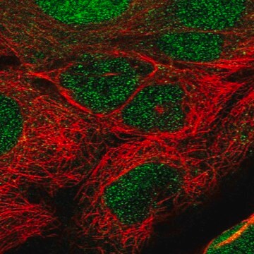
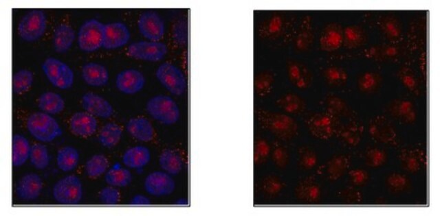
immunofluorescence: 0.25-2 μg/mL
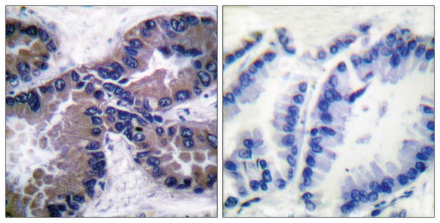
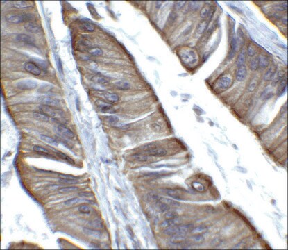
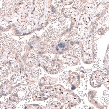
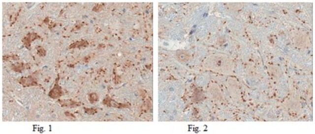
immunohistochemistry: 1:50-1:200
면역원 서열
VPPNPIATFNAPSKWPEPQSTVSYGLAVQGAIQILPLGSGHTPQSSSNSEKNSLPPVMAISNVENEKQVHISFLPANTQGFPLAPERGLFHASLGIAQLSQAGPSKSDRGSSQVSVTSTVHVVNTTVVTMPVPMVSTSSSSYTT
UniProt 수납 번호
배송 상태
wet ice
저장 온도
−20°C
타겟 번역 후 변형
unmodified
유전자 정보
human ... TET1(80312)
일반 설명
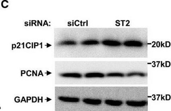
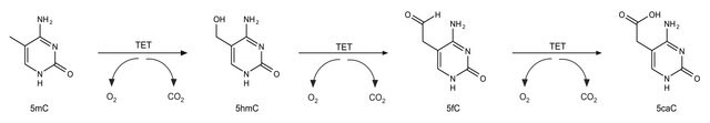
TET1 is a protein that belongs to the TET family and contains a zinc-binding CXXC domain. TET1 functions as a 5-methylcytosine (5mC) hydroxylase and converts 5-methylcytosine to 5-hydroxymethylcytosine which is crucial for active DNA demethylation in the adult brain. Anti-TET1 antibody can be used in western blotting. Rabbit anti-TET1 antibody reacts specifically with TET1 protein.
면역원
Protein TET1 (Ten-eleven translocation 1 gene protein) (CXXC-type zinc finger protein 6) (Leukemia-associated protein with a CXXC domain)
애플리케이션
All Prestige Antibodies Powered by Atlas Antibodies are developed and validated by the Human Protein Atlas (HPA) project and as a result, are supported by the most extensive characterization in the industry.
The Human Protein Atlas project can be subdivided into three efforts: Human Tissue Atlas, Cancer Atlas, and Human Cell Atlas. The antibodies that have been generated in support of the Tissue and Cancer Atlas projects have been tested by immunohistochemistry against hundreds of normal and disease tissues and through the recent efforts of the Human Cell Atlas project, many have been characterized by immunofluorescence to map the human proteome not only at the tissue level but now at the subcellular level. These images and the collection of this vast data set can be viewed on the Human Protein Atlas (HPA) site by clicking on the Image Gallery link. We also provide Prestige Antibodies® protocols and other useful information.
The Human Protein Atlas project can be subdivided into three efforts: Human Tissue Atlas, Cancer Atlas, and Human Cell Atlas. The antibodies that have been generated in support of the Tissue and Cancer Atlas projects have been tested by immunohistochemistry against hundreds of normal and disease tissues and through the recent efforts of the Human Cell Atlas project, many have been characterized by immunofluorescence to map the human proteome not only at the tissue level but now at the subcellular level. These images and the collection of this vast data set can be viewed on the Human Protein Atlas (HPA) site by clicking on the Image Gallery link. We also provide Prestige Antibodies® protocols and other useful information.
특징 및 장점
Prestige Antibodies® are highly characterized and extensively validated antibodies with the added benefit of all available characterization data for each target being accessible via the Human Protein Atlas portal linked just below the product name at the top of this page. The uniqueness and low cross-reactivity of the Prestige Antibodies® to other proteins are due to a thorough selection of antigen regions, affinity purification, and stringent selection. Prestige antigen controls are available for every corresponding Prestige Antibody and can be found in the linkage section.
Every Prestige Antibody is tested in the following ways:
Every Prestige Antibody is tested in the following ways:
- IHC tissue array of 44 normal human tissues and 20 of the most common cancer type tissues.
- Protein array of 364 human recombinant protein fragments.
결합
Corresponding Antigen APREST74805
물리적 형태
Solution in phosphate-buffered saline, pH 7.2, containing 40% glycerol and 0.02% sodium azide
법적 정보
Prestige Antibodies is a registered trademark of Merck KGaA, Darmstadt, Germany
면책조항
Unless otherwise stated in our catalog or other company documentation accompanying the product(s), our products are intended for research use only and are not to be used for any other purpose, which includes but is not limited to, unauthorized commercial uses, in vitro diagnostic uses, ex vivo or in vivo therapeutic uses or any type of consumption or application to humans or animals.
적합한 제품을 찾을 수 없으신가요?
당사의 제품 선택기 도구.을(를) 시도해 보세요.
Storage Class Code
10 - Combustible liquids
WGK
WGK 1
Flash Point (°F)
Not applicable
Flash Point (°C)
Not applicable
개인 보호 장비
Eyeshields, Gloves, multi-purpose combination respirator cartridge (US)
Elham Barazeghi et al.
Clinical epigenetics, 8, 31-31 (2016-03-15)
Primary hyperparathyroidism is characterized by enlarged parathyroid glands due to an adenoma (80-85 %) or multiglandular disease (~15 %) causing hypersecretion of parathyroid hormone (PTH) and generally hypercalcemia. Parathyroid cancer is rare (<1-5 %). The epigenetic mark 5-hydroxymethylcytosine (5hmC) is reduced in various
Sarah Teuber-Hanselmann et al.
International journal of molecular sciences, 22(8) (2021-05-01)
Quantifying O6-methylguanine-DNA methyltransferase (MGMT) promoter methylation plays an essential role in assessing the potential efficacy of alkylating agents in the chemotherapy of malignant gliomas. MGMT promoter methylation is considered to be a characteristic of subgroups of certain malignancies but has
Fu Zhao et al.
Frontiers in oncology, 11, 603686-603686 (2021-03-16)
Medulloblastoma, as the most common malignant brain tumor in children, exhibits highly dysregulated DNA methylation. The novel epigenetic marker-5-hydroxymethylcytosine (5hmC) plays essential role in gene regulation during brain development and in brain tumors. However, the biological and clinical implications of
Mamta Tahiliani et al.
Science (New York, N.Y.), 324(5929), 930-935 (2009-04-18)
DNA cytosine methylation is crucial for retrotransposon silencing and mammalian development. In a computational search for enzymes that could modify 5-methylcytosine (5mC), we identified TET proteins as mammalian homologs of the trypanosome proteins JBP1 and JBP2, which have been proposed
Ryoichi Ono et al.
Cancer research, 62(14), 4075-4080 (2002-07-19)
There are a limited number of reports of acute myeloid leukemia (AML) with t(10;11)(q22;q23). We showed that the MLL gene on 11q23 was fused to the LCX (leukemia-associated protein with a CXXC domain) gene on 10q22 in a de novoadult
자사의 과학자팀은 생명 과학, 재료 과학, 화학 합성, 크로마토그래피, 분석 및 기타 많은 영역을 포함한 모든 과학 분야에 경험이 있습니다..
고객지원팀으로 연락바랍니다.