
추천 제품


생물학적 소스
rabbit
Quality Level
결합
unconjugated
항체 형태
affinity isolated antibody
항체 생산 유형
primary antibodies
클론
polyclonal
제품 라인
Prestige Antibodies® Powered by Atlas Antibodies
양식
buffered aqueous glycerol solution
종 반응성
human
향상된 검증
orthogonal RNAseq
Learn more about Antibody Enhanced Validation
기술
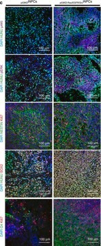
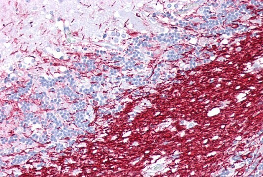
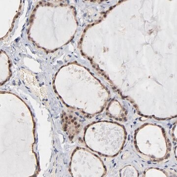
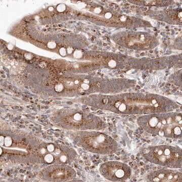
immunohistochemistry: 1:500-1:1000
면역원 서열
LLLAEGFYTTGAVRQIFGDYKTTICGKGLSATVTGGQKGRGSRGQHQAHSLERVCHCLGKWLGHPDKFVGI
UniProt 수납 번호
배송 상태
wet ice
저장 온도
−20°C
타겟 번역 후 변형
unmodified
유전자 정보
human ... PLP1(5354)
일반 설명
Proteolipid protein 1 (PLP1) gene encodes for two major proteins of the central nervous system (CNS) myelin, such as proteolipid protein (PLP) and DM20. This gene is located on the human chromosome at Xq22.2.
면역원
Myelin proteolipid protein recombinant protein epitope signature tag (PrEST)
생화학적/생리학적 작용
Proteolipid protein (PLP) is involved in membrane adhesion, compaction of myelin and synthesis of myelin intra-period line. It is also involved in the maturation of oligodendrocytes. PLP plays a role in wrapping, maintenance and survival of axons. Mutations in the proteolipid protein 1 (PLP1) gene is associated with Pelizaeus-Merzbacher disease indicated by nystagmus, spasticity, microcephaly, ataxia and intellectual disability. PLP1 gene plays a role in Xq22.2 microdeletion and microduplication syndromes. Duplication of this gene results in the overexpression of PLP, which leads to accumulation in the cytoplasm myelinating oligodendrocytes and defects in myelin.
특징 및 장점
Prestige Antibodies® are highly characterized and extensively validated antibodies with the added benefit of all available characterization data for each target being accessible via the Human Protein Atlas portal linked just below the product name at the top of this page. The uniqueness and low cross-reactivity of the Prestige Antibodies® to other proteins are due to a thorough selection of antigen regions, affinity purification, and stringent selection. Prestige antigen controls are available for every corresponding Prestige Antibody and can be found in the linkage section.
Every Prestige Antibody is tested in the following ways:
Every Prestige Antibody is tested in the following ways:
- IHC tissue array of 44 normal human tissues and 20 of the most common cancer type tissues.
- Protein array of 364 human recombinant protein fragments.
결합
Corresponding Antigen APREST74534
물리적 형태
Solution in phosphate-buffered saline, pH 7.2, containing 40% glycerol and 0.02% sodium azide
법적 정보
Prestige Antibodies is a registered trademark of Merck KGaA, Darmstadt, Germany
면책조항
Unless otherwise stated in our catalog or other company documentation accompanying the product(s), our products are intended for research use only and are not to be used for any other purpose, which includes but is not limited to, unauthorized commercial uses, in vitro diagnostic uses, ex vivo or in vivo therapeutic uses or any type of consumption or application to humans or animals.
적합한 제품을 찾을 수 없으신가요?
당사의 제품 선택기 도구.을(를) 시도해 보세요.
Storage Class Code
10 - Combustible liquids
WGK
WGK 1
Flash Point (°F)
Not applicable
Flash Point (°C)
Not applicable
개인 보호 장비
Eyeshields, Gloves, multi-purpose combination respirator cartridge (US)
가장 최신 버전 중 하나를 선택하세요:
Ravinder Pannu et al.
Journal of neuroscience research, 79(3), 340-350 (2004-12-18)
Spinal cord injury (SCI) is a devastating and complex clinical condition involving proinflammatory cytokines and nitric oxide toxicity that produces a predictable pattern of progressive injury entailing neuronal loss, axonal destruction, and demyelination at the site of impact. The involvement
Derek P Ng et al.
Biochemistry, 53(23), 3747-3757 (2014-05-27)
Central to the formation of tertiary structure in membrane protein folding is the presence of amino acid sequence motifs (such as "small-XXX-small" segments) in the TM segments that promote interaction-compatible surfaces through which the TM α-helices interact. Here, we sought
P Martínez-Montero et al.
Clinical genetics, 84(6), 566-571 (2013-01-26)
Pelizaeus-Merzbacher disease (PMD) is caused in most cases by either duplications or point mutations in the PLP1 gene. This disease, a dysmyelinating disorder affecting mainly the central nervous system, has a wide clinical spectrum and its causing mutations act through
Dorota Hoffman-Zacharska et al.
Medycyna wieku rozwojowego, 17(4), 293-300 (2014-02-13)
The Pelizaeus-Merzbacher disease (PMD) is a rare X-linked recessive hypomyelination disorder caused by mutations of the proteolipid protein1 gene (PLP1). There is a spectrum of PLP1-related disorders from very severe connatal PMD, through classical PMD to mild spastic paraplegia type
A microdeletion at Xq22. 2 implicates a glycine receptor GLRA4 involved in intellectual disability, behavioral problems and craniofacial anomalies
Labonne J D J, et al.
BMC Neurology, 16(1), 1-12 (2016)
자사의 과학자팀은 생명 과학, 재료 과학, 화학 합성, 크로마토그래피, 분석 및 기타 많은 영역을 포함한 모든 과학 분야에 경험이 있습니다..
고객지원팀으로 연락바랍니다.