
추천 제품


제품명
Monoclonal Anti-Dystrophin antibody produced in mouse, clone MANDRA1, ascites fluid
생물학적 소스
mouse
결합
unconjugated
항체 형태
ascites fluid
항체 생산 유형
primary antibodies
클론
MANDRA1, monoclonal
포함
15 mM sodium azide
종 반응성
rat, human, mouse, fish
기술
indirect immunofluorescence: 1:100 using freshly dissected and frozen human or animal muscle tissue.
microarray: suitable
동형
IgG1
UniProt 수납 번호
배송 상태
dry ice
저장 온도
−20°C
타겟 번역 후 변형
unmodified
유전자 정보
human ... DMD(1756)
mouse ... Dmd(13405)
rat ... Dmd(24907)
일반 설명
Dystrophin is a muscle membrane protein (427 kDa) which is absent, reduced or altered as a result of mutation in Duchenne and Becker muscular dystrophies (DMD/BMD) or its homologue in the mouse. Severe DMD is associated with a marked dystrophin deficiency whereas patients with the milder form of DMD show less pronounced abnormalities of protein expression. Because abnormalities in the protein expression occur specifically in patients with these types of muscular dystrophy, dystrophin analysis may be used to distinguish these conditions from other neuromuscular diseases. Predictions from the sequence suggest a structural protein on the inner face of the membrane, consisting of a 25-repeat, rod-like triple-helical domain separating an N-terminal actin-binding domain from two C-terminal domains, one of which is rich in cysteine. The large size of dystrophin and its low abundance (<0.01% of the total muscle protein) are a hindrance to the isolation of intact, native protein for structure/function studies.
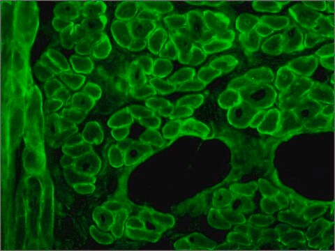
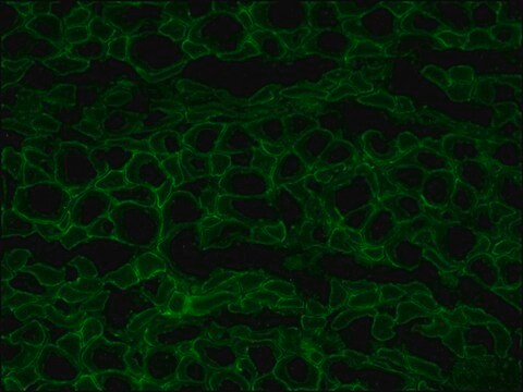
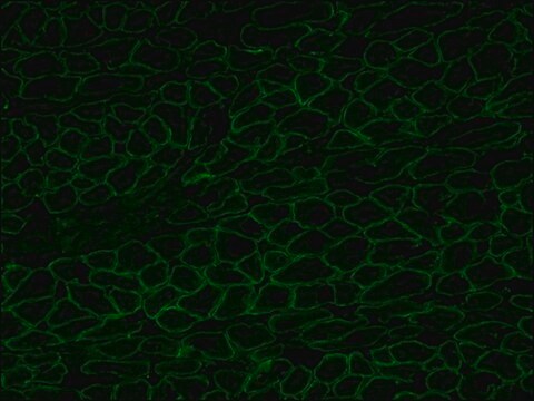
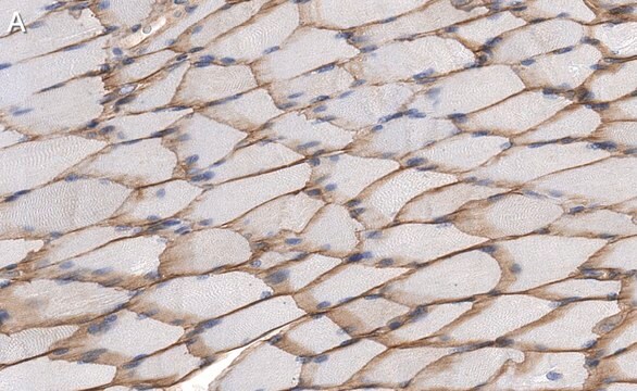
The antibody recognizes an epitope located on the 128 amino acids at the end of the C-terminal domain of the human dystrophin molecule (amino acid residues 3558-3684). Immunohistochemical staining of muscle tissue results in a clear labeling confined to the periphery (plasma membrane) of normal striated muscle fibers. By immunoblotting, the antibody stains dystrophin (427 kDa) in muscle and brain extracts. It also stains the 70-75 kDa protein known as Apo-Dystrophin-1 or Dp71.4 This is detected in the brain as well as in lymphoblastoid cells, cultures of brain astroglial and neuronal cells, liver and Hep G2 cells (human hepatoma). The epitope recognized by the antibody is sensitive to formalin fixation and paraffin embedding. The antibody exhibits a wide interspecies cross-reactivity. It is useful in ELISA and capture ELISA. The antibody is specific to dystrophin and does not react with α-actinin and utrophin, an autosomal homologue of dystrophin, also called dystrophin-related protein (DRP).
특이성
The antibody recognizes an epitope located on the 128 amino acids at the end of the C-terminal domain of the human dystrophin molecule (amino acid residues 3558-3684). Immunohistochemical staining of muscle tissue results in a clear labeling confined to the periphery (plasma membrane) of normal striated muscle fibers. By immunoblotting, the antibody stains dystrophin (427 kDa) in muscle and brain extracts. It also stains the 70-75 kDa protein known as Apo-Dystrophin-1 or Dp71.4 This is detected in the brain as well as in lymphoblastoid cells, cultures of brain astroglial and neuronal cells, liver and Hep G2 cells (human hepatoma). The epitope recognized by the antibody is sensitive to formalin fixation and paraffin embedding. The antibody exhibits a wide interspecies cross-reactivity. It is useful in ELISA and capture ELISA. The antibody is specific to dystrophin and does not react with α-actinin and utrophin, an autosomal homologue of dystrophin, also called dystrophin-related protein (DRP).
면역원
fusion protein containing the C-terminal amino acids of human dystrophine.
애플리케이션
Applications in which this antibody has been used successfully, and the associated peer-reviewed papers, are given below.
Immunofluorescence (1 paper)
Western Blotting (1 paper)
Immunofluorescence (1 paper)
Western Blotting (1 paper)
Mouse monoclonal clone MANDRA1 anti-Dystrophin antibody may be used for the localization of dystrophin using various immunochemical assays such as ELISA, capture ELISA, immunoblot, and immunohistochemistry. Monoclonal antibodies against defined regions of dystrophin provide a means for studying its structure and function, interactions with other proteins and the nature of the partial gene products produced in some patients carrying deletions in the dystrophin gene. The antibodies are useful in the prenatal or post-abortion diagnosis of muscular dystrophy carriers by immunohistological analyses.
표적 설명
The C-terminal domain of the human dystrophin molecule (amino acids residues 3558-3684) is present in normal muscle tissue. It is also present in nearly all Becker muscular dystrophies, but is absent in cases of Duchenne muscular dystrophies and in the dystrophic mouse (mdx).
면책조항
Unless otherwise stated in our catalog or other company documentation accompanying the product(s), our products are intended for research use only and are not to be used for any other purpose, which includes but is not limited to, unauthorized commercial uses, in vitro diagnostic uses, ex vivo or in vivo therapeutic uses or any type of consumption or application to humans or animals.
적합한 제품을 찾을 수 없으신가요?
당사의 제품 선택기 도구.을(를) 시도해 보세요.
Storage Class Code
13 - Non Combustible Solids
WGK
WGK 1
Flash Point (°F)
Not applicable
Flash Point (°C)
Not applicable
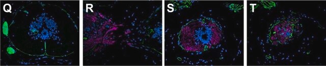
Julia Saifetiarova et al.
Journal of neuroscience research, 95(7), 1373-1390 (2017-04-04)
Bidirectional interactions between neurons and myelinating glial cells result in formation of axonal domains along myelinated fibers. Loss of axonal domains leads to detrimental consequences on nerve structure and function, resulting in reduced conductive properties and the diminished ability to
Kazumi Zushi et al.
Journal of the Japanese Physical Therapy Association = Rigaku ryoho, 15(1), 1-8 (2012-01-01)
The purpose of this study was to investigate the effect of reloading on atrophied muscle and the time course of hypertrophy and regeneration. Forty-nine male Wistar rats were randomly assigned to groups for hindlimb suspension (HS), hindlimb suspension and reloading
Michelle Goody et al.
PLoS currents, 9 (2017-12-01)
Both genetic and infectious diseases can result in skeletal muscle degeneration, inflammation, pain, and/or weakness. Duchenne muscular dystrophy (DMD) is the most common congenital muscle disease. DMD causes progressive muscle wasting due to mutations in Dystrophin. Influenza A and B
Tomonari Awaya et al.
PloS one, 7(12), e51638-e51638 (2012-12-14)
Human embryonic stem (ES) cells and induced pluripotent stem (iPS) cells are promising sources for the cell therapy of muscle diseases and can serve as powerful experimental tools for skeletal muscle research, provided an effective method to induce skeletal muscle
Rasha Al-Khalidi et al.
Acta neuropathologica communications, 6(1), 27-27 (2018-04-13)
Duchenne muscular dystrophy (DMD) is the most common inherited muscle disorder that causes severe disability and death of young men. This disease is characterized by progressive muscle degeneration aggravated by sterile inflammation and is also associated with cognitive impairment and
자사의 과학자팀은 생명 과학, 재료 과학, 화학 합성, 크로마토그래피, 분석 및 기타 많은 영역을 포함한 모든 과학 분야에 경험이 있습니다..
고객지원팀으로 연락바랍니다.