
추천 제품


생물학적 소스
mouse
Quality Level
100
500
결합
unconjugated
항체 형태
culture supernatant
항체 생산 유형
primary antibodies
클론
HMB-45+A103+T311, monoclonal
설명
For In Vitro Diagnostic Use in Select Regions (See Chart)
양식
buffered aqueous solution
종 반응성
human
포장
bottle of 1.0 mL predilute (904H-07)
bottle of 7.0 mL predilute (904H-08)
제조업체/상표
Cell Marque®
동형
IgG1κ
IgG1
IgG2a
배송 상태
wet ice
저장 온도
2-8°C
일반 설명
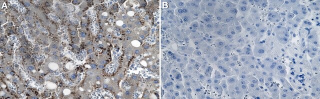
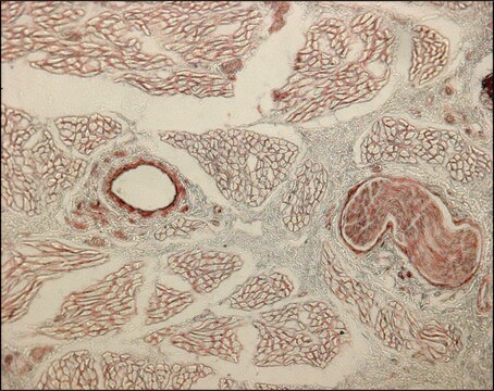
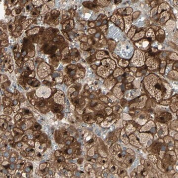
Anti-HMB-45 identifies immature melanosomes. It shows greater specificity for malignant melanomas and metastatic melanomas. MART-1 (also known as Melan A) is a melanocyte differentiation antigen. It is present in melanocytes of normal skin and retina, nevi and in more than 85% of melanomas. Tyrosinase is an enzyme integral in the process of melanin synthesis, and found in 85% to 90% of malignant melanomas. Given these statistics, this cocktail is ideally suited to detection of melanomas and melanocytic lesions.
품질
 IVD |  IVD |  IVD |  RUO |
물리적 형태
Solution in Tris Buffer, pH 7.3-7.7, with 1% BSA and <0.1% Sodium Azide
제조 메모
Download the IFU specific to your product lot and formatNote: This requires a keycode which can be found on your packaging or product label.
기타 정보
For Technical Service please contact: 800-665-7284 or email: service@cellmarque.com
법적 정보
Cell Marque is a registered trademark of Merck KGaA, Darmstadt, Germany
적합한 제품을 찾을 수 없으신가요?
당사의 제품 선택기 도구.을(를) 시도해 보세요.
Storage Class Code
12 - Non Combustible Liquids
WGK
WGK 1
Flash Point (°F)
Not applicable
Flash Point (°C)
Not applicable
가장 최신 버전 중 하나를 선택하세요:
시험 성적서(COA)
Lot/Batch Number
M L Prasad et al.
The American journal of surgical pathology, 25(6), 782-787 (2001-06-08)
Malignant melanomas of the oral and sinonasal mucosa are rare tumors. Amelanotic variants can, on occasion, be difficult to recognize by routine light microscopy. Immunohistochemical studies may be needed for a final diagnosis. A number of new monoclonal antibodies to
Vinod B Shidham et al.
BMC cancer, 3, 15-15 (2003-05-09)
MART-1, Melan-A, and Tyrosinase have shown encouraging results for evaluation of melanoma micrometastases in sentinel lymph nodes, as compared to conventionally used S-100 protein and HMB-45. To achieve higher sensitivity, some studies recommend evaluation of three sections, each at intervals
B L Baisden et al.
The American journal of surgical pathology, 24(8), 1140-1146 (2000-08-10)
Despite the profound therapeutic and prognostic implications of nodal metastases in patients with melanoma, there is no consensus strategy for the optimal detection of metastases in sentinel lymph node biopsies. Traditional microscopic examination may be too crude to detect scattered
L Vaggelli et al.
Tumori, 86(4), 346-348 (2000-10-04)
Elective lymph node dissection (ELND) for patients with malignant melanoma is still controversial. A possible alternative could be biopsy of the first tumor draining lymph node, the sentinel node (SN), which can be identified by means of radionuclide techniques. Our
Deepali Gupta et al.
The American journal of surgical pathology, 26(11), 1450-1457 (2002-11-01)
Malignant melanomas of the vagina are rare tumors. In this study we present the clinicopathologic features and immunohistochemical analysis of 26 such cases seen in our institution over a period of 30 years. The patients' age ranged from 38 to
문서
Immunohistochemistry (IHC) techniques and applications have greatly improved, dermatopathology is still largely based on H&E stained slides.This paper outlines ways in which IHC antibodies can be utilized for dermatopathology.
자사의 과학자팀은 생명 과학, 재료 과학, 화학 합성, 크로마토그래피, 분석 및 기타 많은 영역을 포함한 모든 과학 분야에 경험이 있습니다..
고객지원팀으로 연락바랍니다.