추천 제품
생물학적 소스
rabbit
Quality Level
100
500
결합
unconjugated
항체 형태
Ig fraction of antiserum
항체 생산 유형
primary antibodies
클론
polyclonal
설명
For In Vitro Diagnostic Use in Select Regions (See Chart)
양식
buffered aqueous solution
종 반응성
human
포장
vial of 0.1 mL concentrate (369A-14)
vial of 0.5 mL concentrate (369A-15)
bottle of 1.0 mL predilute (369A-17)
vial of 1.0 mL concentrate (369A-16)
bottle of 7.0 mL predilute (369A-18)
제조업체/상표
Cell Marque®
기술
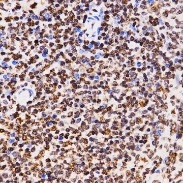
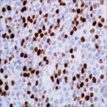
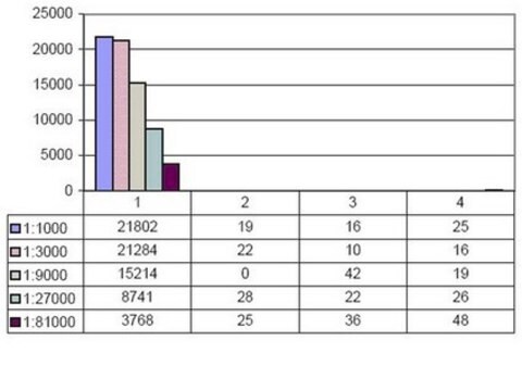
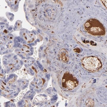
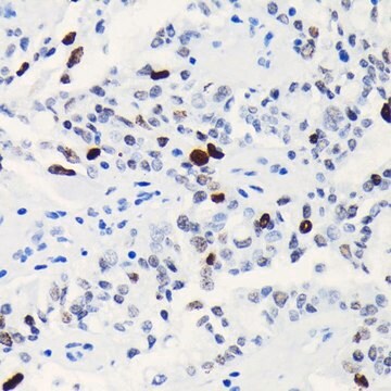
immunohistochemistry (formalin-fixed, paraffin-embedded sections): 1:100-1:500
제어
tonsil
배송 상태
wet ice
저장 온도
2-8°C
시각화
nuclear
유전자 정보
human ... H3C1(8350)
일반 설명
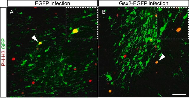
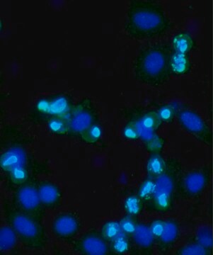
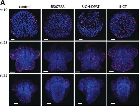
Phosphohistone H3 (PHH3) is a core histone protein, which together with other histones, forms the major protein constituents of the chromatin in eukaryotic cells. In mammalian cells, phosphohistone H3 is negligible during interphase but reaches a maximum for chromatin condensation during mitosis. Immunohistochemical studies showed anti-PHH3 specifically detected the core protein histone H3 only when phosphorylated at serine 10 or serine 28. Studies have also revealed no phosphorylation on the histone H3 during apoptosis. Therefore, PHH3 can serve as a mitotic marker to separate mitotic figures from apoptotic bodies and karyorrhectic debris, which may be a very useful tool in the diagnosis of tumor grades, especially in CNS, skin, gyn., soft tissue, and GIST.
품질
 IVD |  IVD |  IVD |  RUO |
결합
Phosphohistone H3 (PHH3) Positive Control Slides, Product No. 369S, are available for immunohistochemistry (formalin-fixed, paraffin-embedded sections).
물리적 형태
Solution in Tris Buffer, pH 7.3-7.7, with 1% BSA and <0.1% Sodium Azide
제조 메모
Download the IFU specific to your product lot and formatNote: This requires a keycode which can be found on your packaging or product label.
기타 정보
For Technical Service please contact: 800-665-7284 or email: service@cellmarque.com
법적 정보
Cell Marque is a registered trademark of Merck KGaA, Darmstadt, Germany
적합한 제품을 찾을 수 없으신가요?
당사의 제품 선택기 도구.을(를) 시도해 보세요.
가장 최신 버전 중 하나를 선택하세요:
시험 성적서(COA)
Lot/Batch Number
M J Hendzel et al.
The Journal of biological chemistry, 273(38), 24470-24478 (1998-09-12)
Apoptosis plays an important role in the survival of an organism, and substantial work has been done to understand the signaling pathways that regulate this process. Characteristic changes in chromatin organization accompany apoptosis and are routinely used as markers for
Histone phosphorylation and chromatin structure during mitosis in Chinese hamster cells.
L R Gurley et al.
European journal of biochemistry, 84(1), 1-15 (1978-03-01)
Michel R Nasr et al.
The American Journal of dermatopathology, 30(2), 117-122 (2008-03-25)
Differentiating malignant melanoma from benign melanocytic lesions can be challenging. We undertook this study to evaluate the use of the immunohistochemical mitosis marker phospho-Histone H3 (pHH3) and the proliferation markers Ki-67 and survivin in separating malignant melanoma from benign nevi.
Howard Colman et al.
The American journal of surgical pathology, 30(5), 657-664 (2006-05-16)
Distinguishing between grade II and grade III diffuse astrocytomas is important both for prognosis and for treatment decision-making. However, current methods for distinguishing between grades based on proliferative potential are suboptimal, making identification of clear cutoffs difficult. In this study
Yoo-Jin Kim et al.
American journal of clinical pathology, 128(1), 118-125 (2007-06-21)
Mitotic activity is one of the most reliable prognostic factors in meningiomas. The identification of mitotic figures (MFs) and the areas of highest mitotic activity in H&E-stained slides is a tedious and subjective task. Therefore, we compared the results from
자사의 과학자팀은 생명 과학, 재료 과학, 화학 합성, 크로마토그래피, 분석 및 기타 많은 영역을 포함한 모든 과학 분야에 경험이 있습니다..
고객지원팀으로 연락바랍니다.