콘텐츠로 건너뛰기
Merck
모든 사진(1)

문서

B6529

Sigma-Aldrich

Brilliant Blue R Staining Solution

ethanol solution

동의어(들):

Brilliant Blue R, Acid Blue 83, Brilliant indocyanin 6B, Coomassie Brilliant Blue R

로그인조직 및 계약 가격 보기


About This Item

실험식(Hill 표기법):
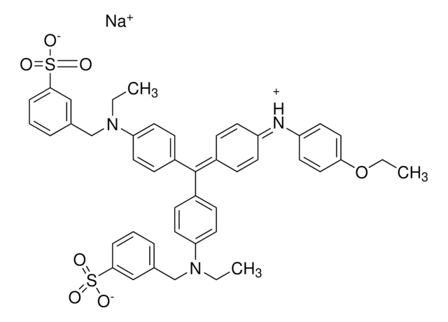
C45H44N3NaO7S2
CAS Number:
Molecular Weight:
825.97
색상 지수 번호:
42660
Beilstein:
5718025
MDL number:
UNSPSC 코드:
12171500
PubChem Substance ID:
NACRES:
NA.47

product name

Brilliant Blue R Staining Solution, suitable for (for immunoelectrophoresis protein staining)

형태

liquid

Quality Level

색상

dark blue

적합성

suitable for (for immunoelectrophoresis protein staining)

응용 분야

diagnostic assay manufacturing
hematology
histology

저장 온도

room temp

SMILES string

[Na+].CCOc1ccc(Nc2ccc(cc2)C(\c3ccc(cc3)N(CC)Cc4cccc(c4)S([O-])(=O)=O)=C5\C=C/C(C=C5)=[N+](\CC)Cc6cccc(c6)S([O-])(=O)=O)cc1

InChI

1S/C45H45N3O7S2.Na/c1-4-47(31-33-9-7-11-43(29-33)56(49,50)51)40-23-15-36(16-24-40)45(35-13-19-38(20-14-35)46-39-21-27-42(28-22-39)55-6-3)37-17-25-41(26-18-37)48(5-2)32-34-10-8-12-44(30-34)57(52,53)54;/h7-30H,4-6,31-32H2,1-3H3,(H2,49,50,51,52,53,54);/q;+1/p-1

InChI key

NKLPQNGYXWVELD-UHFFFAOYSA-M

유사한 제품을 찾으십니까? 방문 제품 비교 안내

애플리케이션

Brilliant Blue R staining solution is especially designed for use in staining protein in agarose gels following immunoelectrophoresis and Ouchterlony gels. The stain contains ethanol and acetic acid so gels do not require fixing prior to staining. The immunoelectrophoresis or Ouchterlony gel is first washed with water and saline to remove non-precipitated proteins and then dried. The gel is then immersed in staining solution for 30 min and destained with 10% acetic acid.
For detection of protein bands following electrophoresis.

성분

0.5% (w/v) Brilliant Blue R, 45% (v/v) ethanol and 10% (v/v) acetic acid.

분석 메모

Tested for suitability on agarose gels following immunoelectrophoresis.

법적 정보

픽토그램

FlameExclamation mark

신호어

Warning

유해 및 위험 성명서

Hazard Classifications

Eye Irrit. 2 - Flam. Liq. 3 - Skin Irrit. 2

Storage Class Code

3 - Flammable liquids

WGK

WGK 2

Flash Point (°F)

81.0 °F - closed cup

Flash Point (°C)

27.2 °C - closed cup

개인 보호 장비

Faceshields, Gloves, Goggles, type ABEK (EN14387) respirator filter


Choose from one of the most recent versions:

시험 성적서(COA)

Lot/Batch Number

Don't see the Right Version?

If you require a particular version, you can look up a specific certificate by the Lot or Batch number.

이 제품을 이미 가지고 계십니까?

문서 라이브러리에서 최근에 구매한 제품에 대한 문서를 찾아보세요.

문서 라이브러리 방문

Wei Zhang et al.
International journal of oncology, 54(5), 1719-1733 (2019-03-14)
Ovarian cancer remains the most lethal type of cancer among all gynecological malignancies. The majority of patients are diagnosed with ovarian cancer at the late stages of the disease. Therefore, there exists an imperative need for the development of early
K Kubo
Analytical biochemistry, 213(2), 200-205 (1993-09-01)
In sodium dodecyl sulfate-polyacrylamide gel electrophoresis (SDS-PAGE) according to the method of Weber and Osborn, protein bands are often distorted by tailing at both ends, and the phenomenon is often called "edge tailing." This was eliminated by adding glycerol at
Zhang Xin-Guo Chen Jian-Hua et al.
Journal of chromatographic science, 50(9), 820-825 (2012-06-22)
A human serum albumin and Thymosin α1 (HSA-Tα1) fusion protein was designed and over-expressed in Pichia pastoris. To purify the fusion protein, a new native preparative electrophoresis system that involved a modified device with a sample receiving chamber, and an
Reiner Westermeier
Proteomics, 6 Suppl 2, 61-64 (2006-10-13)
In spite of the high sensitivity of silver staining and the wide dynamic range of various fluorescent detection methods, Coomassie Brilliant Blue staining is still the most widely used protein detection technique for proteins separated by polyacrylamide gel electrophoresis. There
Chun Yi Liau et al.
Journal of bioscience and bioengineering, 106(1), 111-113 (2008-08-12)
A modified Coomassie Brilliant Blue G 250 staining method for detecting chitinolytic enzymes in chitin-containing polyacrylamide gel electrophoresis (PAGE) is presented. The staining formed achromatic zones at the locations of the migrated enzyme. Using Streptomyces griseus chitinase, we have demonstrated

문서

MISSION® Target ID Library for Human miRNA Target Identification and Discovery;

To meet the great diversity of protein analysis needs, Sigma offers a wide selection of protein visualization (staining) reagents. EZBlue™ and ProteoSilver™, designed specifically for proteomics, also perform impressively in traditional PAGE formats.

자사의 과학자팀은 생명 과학, 재료 과학, 화학 합성, 크로마토그래피, 분석 및 기타 많은 영역을 포함한 모든 과학 분야에 경험이 있습니다..

고객지원팀으로 연락바랍니다.