
추천 제품


사용
sufficient for ≤100 tests
Quality Level
포장
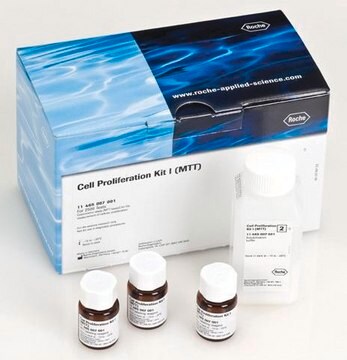
kit of 1 (5 components)
제조업체/상표
Roche
기술
immunofluorescence: suitable
저장 온도
−20°C
일반 설명
The kit is used for the detection of BrdU incorporated into cellular DNA using immunofluorescence microscopy. It is used for the detection of DNA synthesis by either in vitro labeling of cells or organ cultures, or by in vivo labeling, in which frozen or paraffin-embedded tissue sections must be prepared prior to fixation.
Normally, binding of the antibody is only achieved by denaturation of the DNA. This is usually obtained by exposing the cells to acid, base, or heat. These procedures result in destruction of cell integrity, including cell morphology and surface and cytoplasmatic markers.
The BrdU Labeling and Detection Kit I avoids these problems. The antibody preparation contains specific nucleases which allows access to BrdU after fixation in acidic ethanol. Therefore also simultaneous detection of other markers (double staining) is possible.
특이성
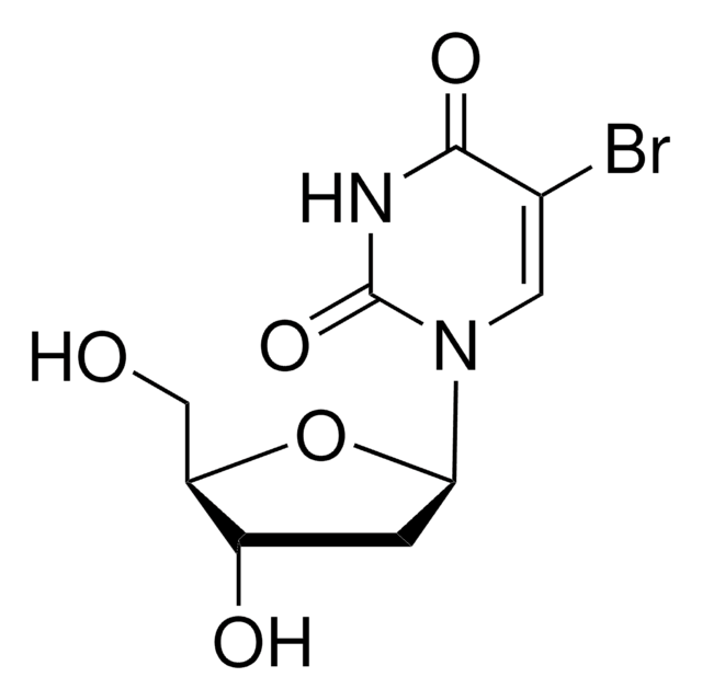
Anti-BrdU monoclonal antibody specifically binds to 5-bromo-2′-deoxy-uridine, and shows cross-reactivity with 5-iodo-2′-deoxy-uridine (10%). Anti-BrdU shows no cross-reactivity with 5-fluoro-2′-deoxy-uridine or any endogenous cellular component, such as thymidine or uridine.
애플리케이션
Samples prelabeled with BrdU are fixed with ethanol, then incubated with a monoclonal antibody to BrdU. The antibody to BrdU supplied with the kit contains an optimized mixture of nucleases. These nucleases generate single-stranded DNA fragments, which allow binding of the antibody to BrdU without destruction of the cellular morphology. A fluorescein-labeled antibody to mouse immunoglobulin is added, then bound to the anti-BrdU antibody. Subsequently, the sample is evaluated using an immunofluorescence microscope.
BrdU Labeling and Detection Kit has been used for the detection of 5-bromo-2′-deoxy-uridine (BrdU) incorporated into cellular DNA.
- Safe: No radioisotopes are used
- Easy to perform: Follows a standard immunofluorescence protocol
- Sensitive: Denaturation of DNA with nucleases allows for highly sensitive detection of BrdU
- Flexible: Allows double-labeling protocols
BrdU Labeling and Detection Kit has been used for the detection of 5-bromo-2′-deoxy-uridine (BrdU) incorporated into cellular DNA.
포장
1 kit containing 5 components.
제조 메모
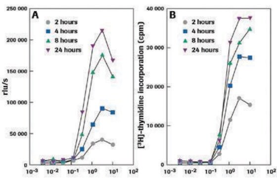
Cell proliferation may be studied by monitoring the incorporation of a radioisotope, [3H]-thymidine, into cellular DNA, followed by autoradiography. Alternatively, 5-bromo-2′-deoxy-uridine (BrdU) may be used instead of thymidine. Cells that have incorporated BrdU into DNA are easily detected using a monoclonal antibody against BrdU and an enzyme- or fluorochrome-conjugated second antibody.
Working solution: BrdU labeling medium
Dilute BrdU labeling reagent 1:1000 with sterile cell culture medium (final concentration 10μM).
Note: For in vivo labeling undiluted BrdU labeling reagent (1 to 2ml/100 g body weight) is needed.
Prepare shortly before use.
Anti-BrdU working solution
Dilute anti-BrdU solution 1:10 with Incubation buffer.
Prepare shortly before use.
Anti-mouse-Ig-fluorescein stock solution
Dissolve anti-mouse-Ig-fluorescein solution in 1ml double-dist. water.
Anti-mouse-Ig-fluorescein working solution
Dilute anti-mouse Ig-fluorescein stock solution 1:10 with PBS. If an extended storage is desired, add BSA (bovine serum albumin), 10 mg/ml.
Prepare shortly before use.
Washing buffer
Dilute Washing buffer concentrate (10x) (bottle 2) 1:10 with double-dist. water.
Storage conditions (working solution): BrdU labeling medium
Store undiluted (1000x) medium in aliquots at -15 to -25°C.
Anti-BrdU working solution
Store undiluted antibody at -15 to -25°C.
Anti-mouse-Ig-fluorescein stock solution
Stable at 2 to 8°C
Washing buffer
Stable at 2 to 8°C
Sample material: Cell culture: adherent cells, suspension cells, organ, or explant cultures. Tissue sections (after in vivo labeling with BrdU).
기타 정보
For life science research only. Not for use in diagnostic procedures.
키트 구성품 전용
제품 번호
설명
- BrdU Labeling Reagent, sterile 1,000x concentrated
- Washing Buffer concentrate 10x concentrated
- Incubation Buffer
- Anti-BrdU antibody, contains nucleases for DNA denaturation
- Anti-mouse-Ig-fluorescein antibody


신호어
Danger
유해 및 위험 성명서
Hazard Classifications
Aquatic Chronic 3 - Eye Irrit. 2 - Muta. 1B - Skin Irrit. 2 - Skin Sens. 1
Storage Class Code
12 - Non Combustible Liquids
WGK
WGK 2
Flash Point (°F)
does not flash
Flash Point (°C)
does not flash
Rosalie E O'Hara et al.
Canadian journal of kidney health and disease, 6, 2054358119871936-2054358119871936 (2019-09-17)
Nephron progenitor cells derived from the metanephric mesenchyme undergo a complex balance of self-renewal and differentiation throughout kidney development to give rise to the mature nephron. Cell proliferation is an important index of progenitor population dynamics. However, accurate and reproducible
Qianqian He et al.
Oncogenesis, 7(8), 62-62 (2018-08-16)
Chromosomal instability (CIN), a high rate of chromosome loss or gain, is often associated with poor prognosis and drug resistance in cancers. Aneuploid, including near-polyploid, cells contain an abnormal number of chromosomes and exhibit CIN. The post-mitotic cell fates following
Sandra Bauer et al.
The Journal of clinical endocrinology and metabolism, 89(2), 812-822 (2004-02-07)
We have tested the hypothesis that elevated concentrations of TNF alpha could impair trophoblast invasion. Using first-trimester placental explant cultures, we have demonstrated that the cytokine inhibits in vitro migration of extravillous trophoblasts (EVT) on collagen I, and invasion through
Yuanyuan Xue et al.
Cell reports, 27(5), 1567-1578 (2019-05-03)
In vertebrates, hematopoiesis occurring in different niches is orchestrated by intrinsic and extrinsic regulators. Previous studies have revealed numerous linear and planar regulatory mechanisms. However, a multi-dimensional transcriptomic atlas of any given hematopoietic organ has not yet been established. Here
M Jesús Gómez-Lamarca et al.
Journal of cell science, 127(Pt 21), 4667-4678 (2014-09-03)
Coordinating exit from the cell cycle with differentiation is crucial for proper development and tissue homeostasis. Failure to do so can lead to aberrant organogenesis and tumorigenesis. However, little is known about the developmental signals that regulate the switch from
문서
Cell based assays for cell proliferation (BrdU, MTT, WST1), cell viability and cytotoxicity experiments for applications in cancer, neuroscience and stem cell research.
자사의 과학자팀은 생명 과학, 재료 과학, 화학 합성, 크로마토그래피, 분석 및 기타 많은 영역을 포함한 모든 과학 분야에 경험이 있습니다..
고객지원팀으로 연락바랍니다.