추천 제품
일반 설명
Rat mammary gland undergoes programmed cell death processes during the tissue regression that follows lactation. Apoptotic bodies, which can be seen by routine histologic staining, can be specifically stained by techniques that localize DNA strand breaks in situ. The specificity of staining for strand breaks can thus be correlated with the morphological changes that define the process of apoptosis.
This product is intended as a positive control for use with all ApopTag In Situ Apoptosis Detection Kits. These reference slides are also useful as teaching aides, and for procedural troubleshooting. Each pack of ApopTag Control Slides contains 5 unstained positive control slides. Rat mammary glands were obtained at the fourth day after weaning and were fixed for 18 hours in 10% neutral buffered formalin. After embedding in paraffin, 5 μm thick sections were cut from the middle of the tissue and were mounted on silanized slides.
애플리케이션
Interpretation of Staining
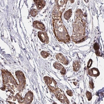
These positive control slides of female rat mammary tissue 4-days post weaning show only 1-2% of the cells being apoptotic. Microscopic inspection is usually required to confirm proper kit activity.
Stain is generally localized to chromatin, and is intensely colored. The localization and sensitivity to be expected with the ApopTag Peroxidase (S7100, S7101) or ApopTag Fluorescein (S7110, S7111, S7160, S7165) Kits are similar. Positive objects vary in size, from intact nuclei to much smaller apoptotic bodies, and in shape, from round to irregular. Such objects may be either isolated, clustered, or engulfed in the cytoplasm of a phagocytic cell. Some unstained apoptotic nuclei or bodies may also be expected. Light cytoplasmic staining might co-localize with intense chromatin staining (this might be due to fragmented DNA leaching from the nucleus, or that in phagocytic organelles). If staining of non-apoptotic cell nuclei is observed with the ApopTag Peroxidase kits (S7100, S7101), the user can attempt to decrease the substrate development time or temperature, or to further dilute the anti-digoxigenin antibody reagent in 0.5% (w:v) BSA. Counterstaining is recommended (see The Complete ApopTag Manual).
Limitations
This control tissue has been selected for positive staining with all ApopTag Kits. Results with control tissue may vary from results with other tissues because of both sample preparation differences and intrinsic tissue differences.
품질
ApopTag Control Slides have been qualified for use with all ApopTag Kits.
저장 및 안정성
Recommended Storage 18°C to 25°C.
법적 정보
CHEMICON is a registered trademark of Merck KGaA, Darmstadt, Germany
면책조항
Unless otherwise stated in our catalog or other company documentation accompanying the product(s), our products are intended for research use only and are not to be used for any other purpose, which includes but is not limited to, unauthorized commercial uses, in vitro diagnostic uses, ex vivo or in vivo therapeutic uses or any type of consumption or application to humans or animals.
시험 성적서(COA)
제품의 로트/배치 번호를 입력하여 시험 성적서(COA)을 검색하십시오. 로트 및 배치 번호는 제품 라벨에 있는 ‘로트’ 또는 ‘배치’라는 용어 뒤에서 찾을 수 있습니다.
Local exposure of 849 MHz and 1763 MHz radiofrequency radiation to mouse heads does not induce cell death or cell proliferation in brain.
Kim, TH; Kim, TH; Huang, TQ; Jang, JJ; Kim, MH; Kim, HJ; Lee, JS; Pack, JK; Seo, JS; Park, WY
Experimental & Molecular Medicine null
자사의 과학자팀은 생명 과학, 재료 과학, 화학 합성, 크로마토그래피, 분석 및 기타 많은 영역을 포함한 모든 과학 분야에 경험이 있습니다..
고객지원팀으로 연락바랍니다.