
S7110
ApopTag Fluorescein In Situ Apoptosis Detection Kit
The ApopTag Fluorescein In Situ Apoptosis Detection Kit detects apoptotic cells in situ by the indirect TUNEL method, utilizing an anti-digoxigenin antibody that is conjugated to a Fluorescein reporter molecule.
동의어(들):
Apoptosis detection kit
로그인조직 및 계약 가격 보기
모든 사진(1)
About This Item
UNSPSC 코드:
12161503
eCl@ss:
32161000
NACRES:
NA.84
추천 제품


Quality Level
제조업체/상표
ApopTag
Chemicon®
기술
flow cytometry: suitable
immunocytochemistry: suitable
immunohistochemistry (formalin-fixed, paraffin-embedded sections): suitable
검출 방법
fluorometric
배송 상태
dry ice
일반 설명
The ApopTag Fluorescein In Situ Apoptosis Detection Kit detects apoptotic cells in situ by the indirect TUNEL method, utilizing an anti-digoxigenin antibody that is conjugated to a Fluorescein reporter molecule. It provides indirect immunofluorescence staining for 40 samples. Results are analyzed using either flow cytometry or fluorescence microscopy.
The ApopTag Fluorescein In Situ Apoptosis Detection Kit has been tested for specific staining in these model systems: (a) human normal peripheral blood lymphocytes induced with dexamethasone as stained in cytospins, (b) rat regressing mammary gland as stained in formalin-fixed, paraffin-embedded sections, and (c) human leukemic peripheral blood lymphocytes induced with camptothecin, as stained in cell suspensions and used for quantitative flow cytometry.
The ApopTag Fluorescein In Situ Apoptosis Detection Kit has been tested for specific staining in these model systems: (a) human normal peripheral blood lymphocytes induced with dexamethasone as stained in cytospins, (b) rat regressing mammary gland as stained in formalin-fixed, paraffin-embedded sections, and (c) human leukemic peripheral blood lymphocytes induced with camptothecin, as stained in cell suspensions and used for quantitative flow cytometry.
애플리케이션
INTRODUCTION
ApopTag In Situ Apoptosis Detection Kits label apoptotic cells in research samples by modifying genomic DNA utilizing terminal deoxynucleotidyl transferase (TdT) for detection of positive cells by specific staining. This manual contains information and protocols for the ApopTag Fluorescein In Situ Apoptosis Detection Kit (Catalog number S7110).
Principles of the Procedure
The reagents provided in all ApopTag Kits are designed to label the free 3′OH DNA termini in situ with chemically labeled and unlabeled nucleotides. The nucleotides contained in the Reaction Buffer are enzymatically added to the DNA by terminal deoxynucleotidyl transferase (TdT) (13, 31). TdT catalyzes a template-independent addition of nucleotide triphosphates to the 3′-OH ends of double-stranded or single-stranded DNA. The incorporated nucleotides form an oligomer composed of digoxigenin nucleotide and unlabeled nucleotide in a random sequence. The ratio of labeled to unlabeled nucleotide in ApopTag Kits is optimized to promote anti-digoxigenin antibody binding, or to minimize fluorescein self-quenching. The exact length of the oligomer added has not been measured.
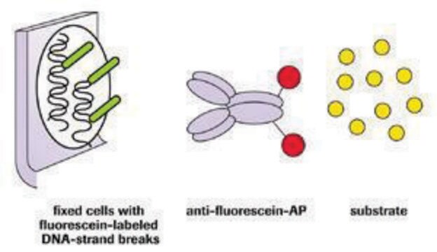
DNA fragments which have been labeled with the digoxigenin-nucleotide are then allowed to bind an anti-digoxigenin antibody that is conjugated to fluorescein (Figure 1A). Fluorescent antibodies provide sensitive detection in immunohistochemistry or immunocytochemistry (i.e. on tissue or cells) and are not subject to experimental variations due to the substrate or the development step. This mixed molecular biological-histochemical systems allows for sensitive and specific staining of very high concentrations of 3′-OH ends that are localized in apoptotic bodies.The ApopTag system differs significantly from previously described in situ labeling techniques for apoptosis (13, 16, 38, 46), in which avidin binding to cellular biotin can be a source of error. The digoxigenin/anti-digoxigenin system has been found to be equally sensitive to avidin/biotin systems (22). Immunochemically-similar ligands for binding of the anti-digoxigenin antibody are generally insignificant in animal tissues, ensuring low background staining. Affinity purified sheep polyclonal antibody is the specific anti-digoxigenin reagent used in ApopTag Kits and exhibits <1% cross-reactivity with the major vertebrate steroids. In addition, the Fc portion of this antibody has been removed by proteolytic digestion to eliminate any non-specific adsorption to cellular Fc receptors.
ApopTag In Situ Apoptosis Detection Kits label apoptotic cells in research samples by modifying genomic DNA utilizing terminal deoxynucleotidyl transferase (TdT) for detection of positive cells by specific staining. This manual contains information and protocols for the ApopTag Fluorescein In Situ Apoptosis Detection Kit (Catalog number S7110).
Principles of the Procedure
The reagents provided in all ApopTag Kits are designed to label the free 3′OH DNA termini in situ with chemically labeled and unlabeled nucleotides. The nucleotides contained in the Reaction Buffer are enzymatically added to the DNA by terminal deoxynucleotidyl transferase (TdT) (13, 31). TdT catalyzes a template-independent addition of nucleotide triphosphates to the 3′-OH ends of double-stranded or single-stranded DNA. The incorporated nucleotides form an oligomer composed of digoxigenin nucleotide and unlabeled nucleotide in a random sequence. The ratio of labeled to unlabeled nucleotide in ApopTag Kits is optimized to promote anti-digoxigenin antibody binding, or to minimize fluorescein self-quenching. The exact length of the oligomer added has not been measured.
DNA fragments which have been labeled with the digoxigenin-nucleotide are then allowed to bind an anti-digoxigenin antibody that is conjugated to fluorescein (Figure 1A). Fluorescent antibodies provide sensitive detection in immunohistochemistry or immunocytochemistry (i.e. on tissue or cells) and are not subject to experimental variations due to the substrate or the development step. This mixed molecular biological-histochemical systems allows for sensitive and specific staining of very high concentrations of 3′-OH ends that are localized in apoptotic bodies.The ApopTag system differs significantly from previously described in situ labeling techniques for apoptosis (13, 16, 38, 46), in which avidin binding to cellular biotin can be a source of error. The digoxigenin/anti-digoxigenin system has been found to be equally sensitive to avidin/biotin systems (22). Immunochemically-similar ligands for binding of the anti-digoxigenin antibody are generally insignificant in animal tissues, ensuring low background staining. Affinity purified sheep polyclonal antibody is the specific anti-digoxigenin reagent used in ApopTag Kits and exhibits <1% cross-reactivity with the major vertebrate steroids. In addition, the Fc portion of this antibody has been removed by proteolytic digestion to eliminate any non-specific adsorption to cellular Fc receptors.
The ApopTag Fluorescein In Situ Apoptosis Detection Kit detects apoptotic cells in situ by the indirect TUNEL method, utilizing an anti-digoxigenin antibody that is conjugated to a Fluorescein reporter molecule.
성분
Equilibration Buffer 90416 3.0 mL -15°C to -25°C
Reaction Buffer 90417 2.0 mL -15°C to -25°C
TdT Enzyme 90418 0.64 mL -15°C to -25°C
Stop/Wash Buffer 90419 20 mL -15°C to -25°C
Blocking Solution 90425 2.6 mL -15°C to -25°C
Anti-Digoxigenin-Fluorescein* 90426 2.1 mL 2°C to 8°C
Plastic Coverslips 90421 100 ea. Room Temp.
*affinity purified sheep polyclonal antibody
저장 및 안정성
1. Store the kit at -15°C to -25°C until the first use. After the first use, if the kit will be used within three months, store the TdT Enzyme (#90418) at -15°C to -25°C and store the remaining components at 2°C to 8°C.
2. Protect the anti-digoxigenin fluorescein antibody (#90426) from unnecessary exposure to light.
Precautions
1. The following kit components contain potassium cacodylate (dimethylarsinic acid) as a buffer: Equilibration Buffer (#90416), Reaction Buffer (#90417), and TdT Enzyme (#90418). These components are harmful if swallowed; avoid contact with skin and eyes (wear gloves, glasses) and wash areas of contact immediately.
2. Antibody Conjugates (#90426) and Blocking Solutions (#90425) contain 0.08% sodium azide as a preservative.
3. TdT Enzyme (#90418) contains glycerol and will not freeze at -20°C. For maximum shelf life, do not warm this reagent to room temperature before dispensing.
법적 정보
CHEMICON is a registered trademark of Merck KGaA, Darmstadt, Germany
면책조항
Unless otherwise stated in our catalog or other company documentation accompanying the product(s), our products are intended for research use only and are not to be used for any other purpose, which includes but is not limited to, unauthorized commercial uses, in vitro diagnostic uses, ex vivo or in vivo therapeutic uses or any type of consumption or application to humans or animals.


신호어
Danger
유해 및 위험 성명서
Hazard Classifications
Aquatic Chronic 2 - Carc. 1B - STOT RE 2 Inhalation
표적 기관
Respiratory Tract
Storage Class Code
6.1C - Combustible acute toxic Cat.3 / toxic compounds or compounds which causing chronic effects
시험 성적서(COA)
제품의 로트/배치 번호를 입력하여 시험 성적서(COA)을 검색하십시오. 로트 및 배치 번호는 제품 라벨에 있는 ‘로트’ 또는 ‘배치’라는 용어 뒤에서 찾을 수 있습니다.
Toru Nakazawa et al.
Molecular vision, 12, 867-878 (2006-08-19)
Photoreceptor apoptosis is associated with retinal detachment (RD) induced photoreceptor degeneration. Previously, we demonstrated the importance of caspase activation for RD-induced photoreceptor death in a rat model of RD. However, extracellular signals that precede the activation of caspases and photoreceptor
Martin Zenker et al.
Nature genetics, 37(12), 1345-1350 (2005-11-29)
Johanson-Blizzard syndrome (OMIM 243800) is an autosomal recessive disorder that includes congenital exocrine pancreatic insufficiency, multiple malformations such as nasal wing aplasia, and frequent mental retardation. We mapped the disease-associated locus to chromosome 15q14-21.1 and identified mutations, mostly truncating ones
Sarah L Lebeis et al.
Journal of immunology (Baltimore, Md. : 1950), 179(1), 566-577 (2007-06-21)
Enteropathogenic Escherichia coli, enterohemorrhagic E. coli, and Citrobacter rodentium are classified as attaching and effacing pathogens based on their ability to adhere to intestinal epithelium via actin-filled membranous protrusions (pedestals). Infection of mice with C. rodentium causes breach of the
Junichiro En et al.
Infection and immunity, 76(5), 2002-2007 (2008-03-05)
Buruli ulcer is a chronic skin disease caused by Mycobacterium ulcerans, which produces a toxic lipid mycolactone. Despite the extensive necrosis and tissue damage, the lesions are painless. This absence of pain prevents patients from seeking early treatment and, as
Brian W Parks et al.
Journal of lipid research, 46(7), 1405-1415 (2005-04-19)
Lysophosphatidylcholine (LPC) is considered a major proatherogenic component of oxidized low density lipoprotein based on its proinflammatory actions in vitro. LPC stimulates macrophage and T-cell chemotaxis via the G protein-coupled receptor G2A and may thus promote inflammatory cell infiltration during
문서
Cellular apoptosis assays to detect programmed cell death using Annexin V, Caspase and TUNEL DNA fragmentation assays.
자사의 과학자팀은 생명 과학, 재료 과학, 화학 합성, 크로마토그래피, 분석 및 기타 많은 영역을 포함한 모든 과학 분야에 경험이 있습니다..
고객지원팀으로 연락바랍니다.