
추천 제품

생물학적 소스
mouse
Quality Level
항체 형태
purified immunoglobulin
항체 생산 유형
primary antibodies
클론
133, monoclonal
종 반응성
rat, human
기술
ELISA: suitable
immunohistochemistry: suitable (paraffin)
neutralization: suitable
western blot: suitable
동형
IgG2bκ
NCBI 수납 번호
UniProt 수납 번호
배송 상태
dry ice
타겟 번역 후 변형
unmodified
유전자 정보
human ... THBS1(7057)
일반 설명
Thrombospondin-1 (UniProt P07996; also known as THBS-1, TSP-1) is encoded by the THBS1 (also known as THBS, TSP, TSP1) gene (Gene ID 7057) in human. Thrombospondin-1 (TSP-1) is a member of the TSP family of calcium-binding extracellular matrix proteins. The TSP family consists of the homotrimeric TSP-1 and TSP-2, as well as the homopentameric TSP-3, TSP-4 and TSP-5/COMP (cartilage oligomeric matrix protein). TSP-1 is induced at injury site and functions as an activator of latent TGF-β. TSP-1 binding alters the conformation of the latent TGF-β complex and renders TGF-β biologically active, which in turn can also induce TSP-1 expression. TSP-1 mediates the proliferation of fibroblasts and smooth muscle cells, while it inhibits the proliferation of endothelial cells. TSP-1 may also serve as both an attachment protein and an anti-adhesive molecule as shown by its ability to cause disassembly of focal adhesions of endothelial cells. TSP-1 is initially produced with a signal peptide sequence (a.a. 1-18), the removal of which yields the mature protein (a.a. 19-1170). Structurally, TSP-1 monomer consists of a N-terminal heparin-binding (a.a. 47-95) region and a laminin G-like domain (a.a. 65-270), followed by an oligomerization domain (a.a. 259-311), a procollagen or VWFC domain (a.a. 316-373), three properdin or type I repeats (a.a. 379-429, 435-490, and 492-547), two EGF-like domains (a.a. 547-587 and 646-690), eight type III repeats or calcium-wire module (a.a. 691-954), and a C-terminal lectin-like globular module or G domain (a.a. 958-170).
특이성
Clone 133 reacted with human thrombospondin-1 (hTSP-1), but not hTSP-2, mouse TSP-1 (mTSP-1), or mTSP-2. Clone 133 detected the C-terminal 50 kDa chymotryptic fragment of stripped TSP as well as recominant fragements containing the Calcium-binding type-3 repeats (a.a. 692-945) region (Annis, D.S., et al. (2006). J. Thromb. Haemost. 4(2):459-468; Schultz-Cherry, S., and Murphy-Ullrich J.E. (1993). J. Cell Biol. 122(4):923-932).
면역원
Alkaline-stripped human platelet thrombospondin-1 (sTSP-1).
Epitope: Calcium-binding type-3 repeats.
애플리케이션
Detect Thrombospondin-1 using this Anti-Thrombospondin-1 Antibody, clone 133 validated for use in Western Blotting, Immunohistochemistry (Paraffin), Neutralizing, ELISA.
Research Category
Cell Structure
Cell Structure
Research Sub Category
ECM Proteins
ECM Proteins
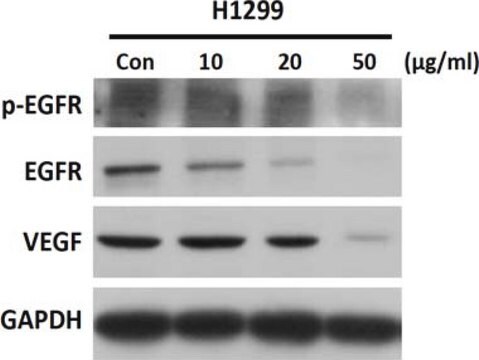
Western Blotting Analysis: A representative lot detected an upregulated thrombospondin-1 (TSP-1) expression in cultured primary rat vascular smooth muscle cells (VSMCs) upon treatment with either stainless steel (SS) ions or active TGF-β (Pallero, M.A., et al. (2010). J. Vasc. Res. 47(4):309-322).
Western Blotting Analysis: A representativev lot ditected full-length human thrombospondin-1 (hTSP-1) as well as hTSP-1 C-terminal fragments E3CaG (a.a. 648-1170), E3Ca (a.a. 648-945) and Ca (a.a. 692-945), but not hTSP-2, mouse TSP-1 (mTSP-1), or mTSP-2 by Western blotting under either reducing or non-reducing condition (Annis, D.S., et al. (2006). J. Thromb. Haemost. 4(2):459-468).
Western Blotting Analysis: A representative lot detected an upregulated thrombospondin-1 (TSP-1) in human pancreatic cancer Panc-1 cells upon siRNA-mediatted k-Ras knockdown (Fleming, J.B., et al. (2005). Mol. Cancer Res. 3(7):413-423).
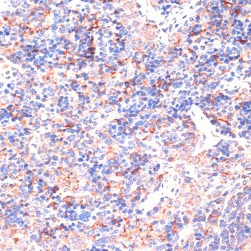
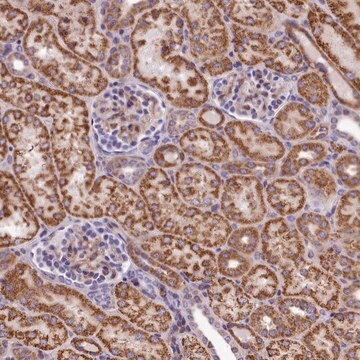
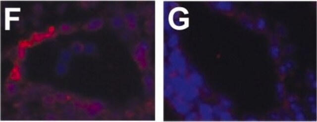
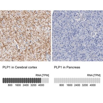
Immunohistochemistry Analysis: A representative lot detected elevated thrombospondin-1 (TSP-1) immunoreactivity in vascular tissues surrounding stent materials due to in-stent restenosis (ISR) in paraffin-embedded sections of human coronary arteries from patients received either Taxus or Cypher drug-eluting stent implant (Pallero, M.A., et al. (2010). J. Vasc. Res. 47(4):309-322).
Neutralizing Analysis: Representative lots prevented alkaline-stripped human platelet thrombospondin-1 (sTSP-1) from activatating the latent form of TGF-beta secreted by cultured NMuMG murine mammary gland epithelial cells or bovine aortic endothelial (BAE) cells without affecting cellular signaling induced by the mature/active form of TGF-beta (Alcaraz, L.B., et al. (2014). J. Cell Biol. 205(3):409-428; Annis, D.S., et al. (2006). J. Thromb. Haemost. 4(2):459-468; Schultz-Cherry, S., and Murphy-Ullrich J.E. (1993). J. Cell Biol. 122(4):923-932).
Neutralizing Analysis: A representative lot inhibited stainless steel (SS) ions-induced TGF-β signaling in cultured primary rat vascular smooth muscle cells (VSMCs) as indicated by a suppressed induction of the synthetic/myofibroblastic protein ED-A FN (Pallero, M.A., et al. (2010). J. Vasc. Res. 47(4):309-322).
ELISA Analysis: A representativev lot ditected full-length human thrombospondin-1 (hTSP-1) as well as hTSP-1 C-terminal fragments E3CaG (a.a. 648-1170) and E3Ca (a.a. 648-945), but not hTSP-2, mouse TSP-1 (mTSP-1), or mTSP-2 by direct/non-sandwich ELISA (Annis, D.S., et al. (2006). J. Thromb. Haemost. 4(2):459-468).
Western Blotting Analysis: A representativev lot ditected full-length human thrombospondin-1 (hTSP-1) as well as hTSP-1 C-terminal fragments E3CaG (a.a. 648-1170), E3Ca (a.a. 648-945) and Ca (a.a. 692-945), but not hTSP-2, mouse TSP-1 (mTSP-1), or mTSP-2 by Western blotting under either reducing or non-reducing condition (Annis, D.S., et al. (2006). J. Thromb. Haemost. 4(2):459-468).
Western Blotting Analysis: A representative lot detected an upregulated thrombospondin-1 (TSP-1) in human pancreatic cancer Panc-1 cells upon siRNA-mediatted k-Ras knockdown (Fleming, J.B., et al. (2005). Mol. Cancer Res. 3(7):413-423).
Immunohistochemistry Analysis: A representative lot detected elevated thrombospondin-1 (TSP-1) immunoreactivity in vascular tissues surrounding stent materials due to in-stent restenosis (ISR) in paraffin-embedded sections of human coronary arteries from patients received either Taxus or Cypher drug-eluting stent implant (Pallero, M.A., et al. (2010). J. Vasc. Res. 47(4):309-322).
Neutralizing Analysis: Representative lots prevented alkaline-stripped human platelet thrombospondin-1 (sTSP-1) from activatating the latent form of TGF-beta secreted by cultured NMuMG murine mammary gland epithelial cells or bovine aortic endothelial (BAE) cells without affecting cellular signaling induced by the mature/active form of TGF-beta (Alcaraz, L.B., et al. (2014). J. Cell Biol. 205(3):409-428; Annis, D.S., et al. (2006). J. Thromb. Haemost. 4(2):459-468; Schultz-Cherry, S., and Murphy-Ullrich J.E. (1993). J. Cell Biol. 122(4):923-932).
Neutralizing Analysis: A representative lot inhibited stainless steel (SS) ions-induced TGF-β signaling in cultured primary rat vascular smooth muscle cells (VSMCs) as indicated by a suppressed induction of the synthetic/myofibroblastic protein ED-A FN (Pallero, M.A., et al. (2010). J. Vasc. Res. 47(4):309-322).
ELISA Analysis: A representativev lot ditected full-length human thrombospondin-1 (hTSP-1) as well as hTSP-1 C-terminal fragments E3CaG (a.a. 648-1170) and E3Ca (a.a. 648-945), but not hTSP-2, mouse TSP-1 (mTSP-1), or mTSP-2 by direct/non-sandwich ELISA (Annis, D.S., et al. (2006). J. Thromb. Haemost. 4(2):459-468).
품질
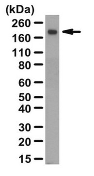
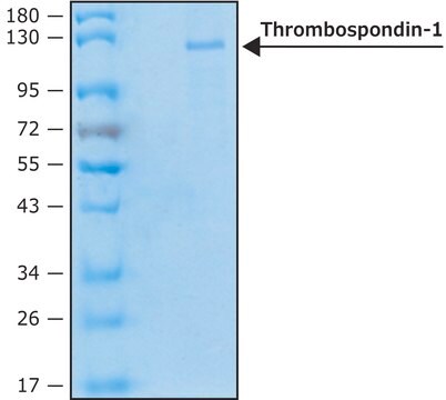
Evaluated by Western Blotting in human thrombospondin-1 purified protein.
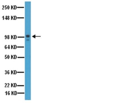
Western Blotting Analysis: 0.1 µg/mL of this antibody detected Thrombospondin-1 in 0.1 µg of human thrombospondin-1 purified protein.
Western Blotting Analysis: 0.1 µg/mL of this antibody detected Thrombospondin-1 in 0.1 µg of human thrombospondin-1 purified protein.
표적 설명
~129 kDa observed.
물리적 형태
Format: Purified
Protein G Purified
Purified mouse monoclonal IgG2bκ antibody in PBS without preservatives.
저장 및 안정성
Stable for 1 year at -20°C from date of receipt.
Handling Recommendations: Upon receipt and prior to removing the cap, centrifuge the vial and gently mix the solution. Aliquot into microcentrifuge tubes and store at -20°C. Avoid repeated freeze/thaw cycles, which may damage IgG and affect product performance.
Handling Recommendations: Upon receipt and prior to removing the cap, centrifuge the vial and gently mix the solution. Aliquot into microcentrifuge tubes and store at -20°C. Avoid repeated freeze/thaw cycles, which may damage IgG and affect product performance.
기타 정보
Concentration: Please refer to lot specific datasheet.
면책조항
Unless otherwise stated in our catalog or other company documentation accompanying the product(s), our products are intended for research use only and are not to be used for any other purpose, which includes but is not limited to, unauthorized commercial uses, in vitro diagnostic uses, ex vivo or in vivo therapeutic uses or any type of consumption or application to humans or animals.
적합한 제품을 찾을 수 없으신가요?
당사의 제품 선택기 도구.을(를) 시도해 보세요.
Storage Class Code
12 - Non Combustible Liquids
WGK
WGK 2
Flash Point (°F)
Not applicable
Flash Point (°C)
Not applicable
시험 성적서(COA)
제품의 로트/배치 번호를 입력하여 시험 성적서(COA)을 검색하십시오. 로트 및 배치 번호는 제품 라벨에 있는 ‘로트’ 또는 ‘배치’라는 용어 뒤에서 찾을 수 있습니다.
자사의 과학자팀은 생명 과학, 재료 과학, 화학 합성, 크로마토그래피, 분석 및 기타 많은 영역을 포함한 모든 과학 분야에 경험이 있습니다..
고객지원팀으로 연락바랍니다.