추천 제품
생물학적 소스
mouse
Quality Level
항체 형태
purified antibody
항체 생산 유형
primary antibodies
클론
1H7-4B, monoclonal
종 반응성
human
기술
ELISA: suitable
flow cytometry: suitable
immunohistochemistry: suitable (paraffin)
western blot: suitable
동형
IgG1κ
NCBI 수납 번호
UniProt 수납 번호
배송 상태
wet ice
타겟 번역 후 변형
unmodified
유전자 정보
human ... CEACAM6(4680)
관련 카테고리
일반 설명
The carcinoembryonic antigen- (CEA-) related cell adhesion molecules (CEACAMs) constitute a 12-member subgroup of the CEA family of immunoglobulin-related proteins first described in 1965 (PMID 4953873). CEACAMs are reported to participate in diverse physiological processes, including cell adhesion, differentiation, proliferation, and survival, as well as in carcinogenesis and bacterial pathogenesis. CEACAM molecules from neighbouring cells can interact via their respective extracellular N-terminal IgV-like domain and mediate cell-cell adhesion through trans-oligomerization. CEACAM molecules within the same cell can also undergo transmembrane domain-mediated cis-oligomerization, an event important for sustaining downstream cellular signaling. Athough CEACAM1 is shown to utilize its cytoplasmic domain for transducing cellular signaling, not all CEACAM members are expressed with a significant cytoplasmic domain, and others (CEACAM5/6/7/8) are GPI-anchored without even a transmembrane domain. (PMID 4735478). Human CEACAM6 (also known as CD66c, Non-specific crossreacting antigen, Normal cross-reacting antigen; UniProt P40199) is encoded by the CEACAM6 (or CEAL & NCA) gene (NCBI Gene ID 4680). It is a GPI-anchored CEACAM with three extracellular Ig domains. Clone 1H7-4B does not cross-react with human CEACAM1/3/4/5/7/8, rat CECAM1, or mouse CEACAM1/2.
특이성
Clone 1H7-4B specifically detected exogenously expressed human CEACAM6, but not CEACAM1/3/4/5/7/8, on the surface of transfected CHO cells.
면역원
Epitope: Extracellular domain.
Recombinant human CEACAM6 extracellular domain.
애플리케이션
Anti-CEACAM6 Antibody, clone 1H7-4B is an antibody against CEACAM6 for use in Western Blotting, Immunohistochemistry (Paraffin), ELISA, Immunoprecipitation, Immunocytochemistry, Flow Cytometry.
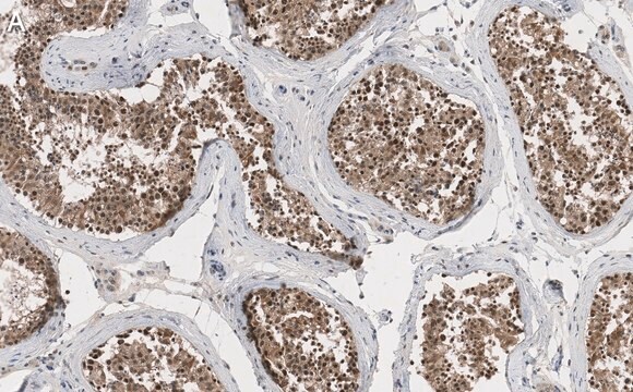
Immunohistochemistry Analysis: A 1:1,000 dilution from a representative lot detected CEACAM6 in normal (colon, lung, lymph node) and cancer (lung, colon) human tissue sections.
Flow Cytometry Analysis: A representative lot detected exogenously expressed human CEACAM6 on the surface of transfected CHO cells (Courtesy of Dr. B. Singer, University Duisburg-Essen, Germany).
ELISA Analysis: Representative lots detected CEACAM6 in multivesicular bodies (MVBs) from 2 day-starved HT29 human epithelial cells and T102/3 human colon epithelial cancer cells, as well as in human bronchoalveolar lavage fluid (BALF) samples (Singer, B.B., et al. (2014). PLoS One. 9(4):e94106; Klaile, E., et al. (2013). Respir. Res. 14:85).
Immunohistochemistry Analysis: A representative lot detected CEACAM6 in paraffin-embedded human lung cancer tissue sections (Klaile, E., et al. (2013) Respir Res. 14:85).
Western Blotting Analysis: A representative lot detected CEACAM6 in 2 day-starved HT29 human epithelial cells and HT29-derived multivesicular bodies (MVBs) (Muturi H.T., et al. (2013) PLoS One. 8(9):e74654).
Research Category
Cell Structure
Cell Structure
Research Sub Category
Adhesion (CAMs)
Adhesion (CAMs)
품질
Evaluated by Western Blotting in HT-29 cell lysate.
Western Blotting Analysis: 0.5 µg/mL of this antibody detected CEACAM6 in 10 µg of HT-29 cell lysate.
Western Blotting Analysis: 0.5 µg/mL of this antibody detected CEACAM6 in 10 µg of HT-29 cell lysate.
표적 설명
~90 kDa observed. Target band size appears larger than the calculated molecular weights of 33.48/37.20 kDa (mature/pro-form) due to high glycosylation.
물리적 형태
Format: Purified
Protein G Purified
Purified mouse IgG1κ in buffer containing 0.1 M Tris-Glycine (pH 7.4), 150 mM NaCl with 0.05% sodium azide.
저장 및 안정성
Stable for 1 year at 2-8°C from date of receipt.
기타 정보
Concentration: Please refer to lot specific datasheet.
면책조항
Unless otherwise stated in our catalog or other company documentation accompanying the product(s), our products are intended for research use only and are not to be used for any other purpose, which includes but is not limited to, unauthorized commercial uses, in vitro diagnostic uses, ex vivo or in vivo therapeutic uses or any type of consumption or application to humans or animals.
적합한 제품을 찾을 수 없으신가요?
당사의 제품 선택기 도구.을(를) 시도해 보세요.
Storage Class Code
12 - Non Combustible Liquids
WGK
WGK 1
Flash Point (°F)
Not applicable
Flash Point (°C)
Not applicable
시험 성적서(COA)
제품의 로트/배치 번호를 입력하여 시험 성적서(COA)을 검색하십시오. 로트 및 배치 번호는 제품 라벨에 있는 ‘로트’ 또는 ‘배치’라는 용어 뒤에서 찾을 수 있습니다.
Bernhard B Singer et al.
PloS one, 9(4), e94106-e94106 (2014-04-20)
Lower respiratory tract bacterial infections are characterized by neutrophilic inflammation in the airways. The carcinoembryonic antigen-related cell adhesion molecule (CEACAM) 8 is expressed in and released by human granulocytes. Our study demonstrates that human granulocytes release CEACAM8 in response to
Esther Klaile et al.
Respiratory research, 14, 85-85 (2013-08-15)
The carcinoembryonic antigen (CEA)-related cell adhesion molecules CEACAM1 (BGP, CD66a), CEACAM5 (CEA, CD66e) and CEACAM6 (NCA, CD66c) are expressed in human lung. They play a role in innate and adaptive immunity and are targets for various bacterial and viral adhesins.
Harrison T Muturi et al.
PloS one, 8(9), e74654-e74654 (2013-09-17)
Normal and malignant cells release a variety of different vesicles into their extracellular environment. The most prominent vesicles are the microvesicles (MVs, 100-1000 nm in diameter), which are shed of the plasma membrane, and the exosomes (70-120 nm in diameter)
자사의 과학자팀은 생명 과학, 재료 과학, 화학 합성, 크로마토그래피, 분석 및 기타 많은 영역을 포함한 모든 과학 분야에 경험이 있습니다..
고객지원팀으로 연락바랍니다.

