
추천 제품


생물학적 소스
mouse
Quality Level
항체 형태
purified antibody
항체 생산 유형
primary antibodies
클론
19A11, monoclonal
분자량
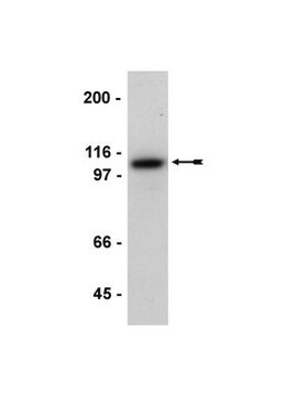
calculated mol wt 97 kDa
정제법
using protein G
종 반응성
human, canine
포장
antibody small pack of 100
기술
ELISA: suitable
immunocytochemistry: suitable
inhibition assay: suitable
western blot: suitable
동형
IgG1κ
에피토프 서열
N-terminal extracellular domain
단백질 ID 수납 번호
UniProt 수납 번호
저장 온도
-10 to -25°C
유전자 정보
human ... CDH1(999)
특이성
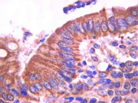
Clone 19A11 is a mouse monoclonal antibody that detects E-Cadherin/CD324. It targets an epitope within the extracellular domain.
면역원
Recombinant protein corresponding to the extracellular domain of human E-Cadherin.
애플리케이션
Quality Control Testing
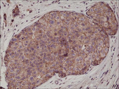
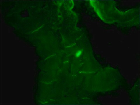
Evaluated by Immunocytochemistry in MCF-7 cells.
Immunocytochemistry Analysis: A 1:1,000 dilution of this antibody detected E-Cadherin/CD324 in MCF-7 cells.
Tested Applications
Agonist Function:: A representative lot stimulated adhesion of cells to pure E-Cadherin substrate (Petrova, Y., et al. (2012). Mol Biol Cell.;23(11):2092-108).
Western Blotting Analysis: A representative lot detected E-Cadherin/CD324) in Western Blotting applications (Petrova, Y., et al. (2012). Mol Biol Cell. 23(11):2092-108; Xie, B., et al. (2022). Proc Natl Acad Sci USA. 119(32):e2204473119).
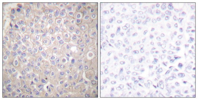
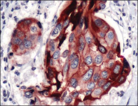
Immunocytochemistry Analysis: A representative lot detected E-Cadherin/CD324 in Immunocytochemistry applications (Petrova, Y., et al. (2012). Mol Biol Cell. 23(11):2092-108).
ELISA Analysis: A representative lot detected E-Cadherin/CD324 in ELISA applications (Petrova, Y., et al. (2012). Mol Biol Cell. 23(11):2092-108).
Note: Actual optimal working dilutions must be determined by end user as specimens, and experimental conditions may vary with the end user.
Evaluated by Immunocytochemistry in MCF-7 cells.
Immunocytochemistry Analysis: A 1:1,000 dilution of this antibody detected E-Cadherin/CD324 in MCF-7 cells.
Tested Applications
Agonist Function:: A representative lot stimulated adhesion of cells to pure E-Cadherin substrate (Petrova, Y., et al. (2012). Mol Biol Cell.;23(11):2092-108).
Western Blotting Analysis: A representative lot detected E-Cadherin/CD324) in Western Blotting applications (Petrova, Y., et al. (2012). Mol Biol Cell. 23(11):2092-108; Xie, B., et al. (2022). Proc Natl Acad Sci USA. 119(32):e2204473119).
Immunocytochemistry Analysis: A representative lot detected E-Cadherin/CD324 in Immunocytochemistry applications (Petrova, Y., et al. (2012). Mol Biol Cell. 23(11):2092-108).
ELISA Analysis: A representative lot detected E-Cadherin/CD324 in ELISA applications (Petrova, Y., et al. (2012). Mol Biol Cell. 23(11):2092-108).
Note: Actual optimal working dilutions must be determined by end user as specimens, and experimental conditions may vary with the end user.
표적 설명
Cadherin-1 (UniProt: P12830; also known as CAM 120/80, Epithelial cadherin, E-cadherin, Uvomorulin, CD324) is encoded by the CDH1 (also known as CDHE, UVO) gene (Gene ID: 999) in human. Cadherins are calcium-dependent cell adhesion molecules that participate in cell-cell adhesion during embryogenesis, development, organogenesis, and differentiation. E-cadherin is a single-pass type I membrane glycoprotein with tumor suppressive properties. It is synthesized with a signal peptide (aa 1-22) and a propeptide (aa 23-154) that are subsequently cleaved off to produce the mature form that contains an extracellular domain (aa 155-709), a transmembrane domain (aa 710-730), and a cytoplasmic domain (aa 731-882). N-glycosylation at Asn-637 is shown to be essential for its expression, folding, and trafficking. E-cadherin is known to contain five cadherin domains. Three calcium ions are usually bound at the interface of each cadherin domain and rigidify the connections, imparting a strong curvature to the full-length ectodomain. Post-translationally it can be cleaved into three chains: E-Cad/CTF1 (aa 701-882); E-Cad/CTF2 (aa 732-882); and E-Cad/CTF3 (aa 751-882). During apoptosis or with calcium influx, it is cleaved by a membrane-bound metalloproteinase (ADAM10; at residues 700-701), which causes disruption of cell-cell adhesion and the subsequent release of β-catenin into the cytoplasm. The residual membrane-tethered cleavage product is then rapidly degraded via an intracellular proteolytic pathway. It can also be cleaved by PS1/ -secretase (at residues 731-732) and this cleavage promotes disassembly of adherens junctions. Clone 19A11 is an activating monoclonal antibody that stimulates adhesion of cells to E-cadherin substrate. It modifies the adhesive properties of E-cadherin and prevent the metastatic invasion of mouse lung cancer cells expressing human E-cadherin. This clone is shown to bind to the extracellular domain of E-cadherin near its primary adhesive motif: the strand swap dimer interface, which strengthens adhesion by stabilizing strand-swap dimers. (Ref.: Xie, B., et al. (2022). Proc. Natl. Acad. Sci. USA. 119 (32); e2204473119; Petrova, YI., et al. (2012). Mol. Biol. Cell. 23(11); 2092-2108; Marambaud, M., et al. (2002). EMBO J. 21(8); 1948-1956; Steinhausen, U., et al. (2001). J. Biol. Chem. 276(7); 4972-4980).
물리적 형태
Purified mouse monoclonal antibody IgG1 in PBS without preservatives.
재구성
0.5 mg/mL. Please refer to guidance on suggested starting dilutions and/or titers per application and sample type.
저장 및 안정성
Store at -10°C to -25°C. Handling Recommendations: Upon receipt and prior to removing the cap, centrifuge the vial and gently mix the solution. Aliquot into microcentrifuge tubes and store at -20°C. Avoid repeated freeze/thaw cycles, which may damage IgG and affect product performance.
기타 정보
Concentration: Please refer to the Certificate of Analysis for the lot-specific concentration.
면책조항
Unless otherwise stated in our catalog or other company documentation accompanying the product(s), our products are intended for research use only and are not to be used for any other purpose, which includes but is not limited to, unauthorized commercial uses, in vitro diagnostic uses, ex vivo or in vivo therapeutic uses or any type of consumption or application to humans or animals.
적합한 제품을 찾을 수 없으신가요?
당사의 제품 선택기 도구.을(를) 시도해 보세요.
Storage Class Code
12 - Non Combustible Liquids
WGK
WGK 2
Flash Point (°F)
Not applicable
Flash Point (°C)
Not applicable
시험 성적서(COA)
제품의 로트/배치 번호를 입력하여 시험 성적서(COA)을 검색하십시오. 로트 및 배치 번호는 제품 라벨에 있는 ‘로트’ 또는 ‘배치’라는 용어 뒤에서 찾을 수 있습니다.
자사의 과학자팀은 생명 과학, 재료 과학, 화학 합성, 크로마토그래피, 분석 및 기타 많은 영역을 포함한 모든 과학 분야에 경험이 있습니다..
고객지원팀으로 연락바랍니다.