
추천 제품
생물학적 소스
mouse
Quality Level
항체 형태
purified antibody
항체 생산 유형
primary antibodies
클론
XB6-AC5, monoclonal
분자량
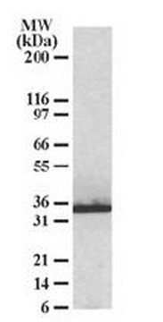
calculated mol wt 41.6 kDa
observed mol wt ~32 kDa
정제법
using protein G
종 반응성
mouse, rat
포장
antibody small pack of 100
기술
immunocytochemistry: suitable
immunofluorescence: suitable
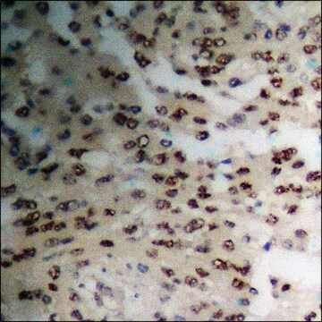
immunohistochemistry: suitable
immunoprecipitation (IP): suitable
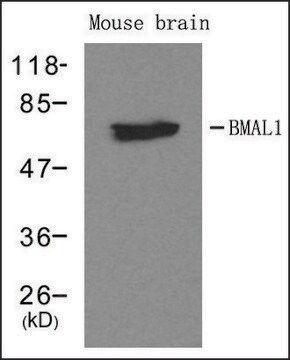
western blot: suitable
동형
IgG1κ
에피토프 서열
N-terminal
단백질 ID 수납 번호
UniProt 수납 번호
저장 온도
2-8°C
유전자 정보
mouse ... Mlc1(170790)
특이성
Clone XB6-AC5 is a mouse monoclonal antibody that detects MLC1. It targets an epitope within 15 amino acids from the N-terminal region.
면역원
A linear peptide corresponding to 15 amino acids from the N-terminal region of mouse MLC1.
애플리케이션
Quality Control Testing
Evaluated by Western Blotting in wild-type Mouse brain membrane extract.
Western Blotting Analysis: A 1:1,000 dilution of this antibody detected MLC1 in Wild-type Mouse brain membrane extract, but not in extract from Mouse membrane with MLC1 knockout.
Tested Applications
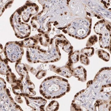
Immunohistochemistry: A representative lot detected MLC1 in Immunohistochemistry applications (Diaz-Castro, B., et al. (2019). Sci Transl Med. 11(514):eaaw8546; Gilbert, A., et al. (2019). Brain Struct Funct. 224(3):1267-1278; Sanchez, A., et al. (2020). Neurotherapeutics. 17(4):2041-2053).
Immunoprecipitation Analysis: Applications: A representative lot detected MLC1 in Immunohistochemistry applications (Elorza-Vidal, X., et al. (2018). Neurobiol Dis. 119:88-99).
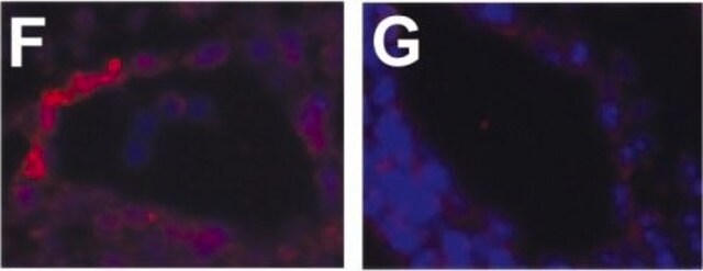
Immunofluorescence Analysis (IF): A representative lot detected MLC1 in Immunofluorescence applications (Sirisi, S., et al. (2017). Hum Mol Genet.;26(13) 2436-2450; Elorza-Vidal, X., et al. (2018). Neurobiol Dis. 119:88-99; Perez-Rius, C., et al. (2019). Orphanet J Rare Dis. 14(1):268; Sanchez, A., et al. (2020). Neurotherapeutics. 17(4):2041-2053).
Immunocytochemistry Analysis: A representative lot detected MLC1 in Immunocytochemistry applications (Sirisi, S., et al. (2017). Hum Mol Genet. 26(13):2436-2450; Elorza-Vidal, X., et al. (2018). Neurobiol Dis. 119:88-99).
Western Blotting Analysis: A representative lot detected MLC1 in Western Blotting applications (Sirisi, S., et al. (2017). Hum Mol Genet. 26(13):2436-2450; Elorza-Vidal, X., et al. (2018). Neurobiol Dis. 119:88-99; Gilbert, A., et al. (2019). Brain Struct Funct. 224(3):1267-1278; Perez-Rius, C., et al. (2019). Orphanet J Rare Dis. 14(1):268; Sanchez, A., et al. (2020). Neurotherapeutics. 17(4):2041-2053).
Note: Actual optimal working dilutions must be determined by end user as specimens, and experimental conditions may vary with the end user.
Evaluated by Western Blotting in wild-type Mouse brain membrane extract.
Western Blotting Analysis: A 1:1,000 dilution of this antibody detected MLC1 in Wild-type Mouse brain membrane extract, but not in extract from Mouse membrane with MLC1 knockout.
Tested Applications
Immunohistochemistry: A representative lot detected MLC1 in Immunohistochemistry applications (Diaz-Castro, B., et al. (2019). Sci Transl Med. 11(514):eaaw8546; Gilbert, A., et al. (2019). Brain Struct Funct. 224(3):1267-1278; Sanchez, A., et al. (2020). Neurotherapeutics. 17(4):2041-2053).
Immunoprecipitation Analysis: Applications: A representative lot detected MLC1 in Immunohistochemistry applications (Elorza-Vidal, X., et al. (2018). Neurobiol Dis. 119:88-99).
Immunofluorescence Analysis (IF): A representative lot detected MLC1 in Immunofluorescence applications (Sirisi, S., et al. (2017). Hum Mol Genet.;26(13) 2436-2450; Elorza-Vidal, X., et al. (2018). Neurobiol Dis. 119:88-99; Perez-Rius, C., et al. (2019). Orphanet J Rare Dis. 14(1):268; Sanchez, A., et al. (2020). Neurotherapeutics. 17(4):2041-2053).
Immunocytochemistry Analysis: A representative lot detected MLC1 in Immunocytochemistry applications (Sirisi, S., et al. (2017). Hum Mol Genet. 26(13):2436-2450; Elorza-Vidal, X., et al. (2018). Neurobiol Dis. 119:88-99).
Western Blotting Analysis: A representative lot detected MLC1 in Western Blotting applications (Sirisi, S., et al. (2017). Hum Mol Genet. 26(13):2436-2450; Elorza-Vidal, X., et al. (2018). Neurobiol Dis. 119:88-99; Gilbert, A., et al. (2019). Brain Struct Funct. 224(3):1267-1278; Perez-Rius, C., et al. (2019). Orphanet J Rare Dis. 14(1):268; Sanchez, A., et al. (2020). Neurotherapeutics. 17(4):2041-2053).
Note: Actual optimal working dilutions must be determined by end user as specimens, and experimental conditions may vary with the end user.
표적 설명
Membrane protein MLC1 (UniProt: Q8VHK5; also known as MLC1) is encoded by the Mlc1 gene (Gene ID: 170790) in murine species. MLC1 is a multi-pass membrane protein that regulates the response of astrocytes to hypo-osmosis by promoting calcium influx. It is a highly hydrophobic protein with eight transmembrane domains and short amino and carboxylic- cytoplasmic tails. It forms highly stable dimeric and oligomeric structures. Within the brain, it is mainly expressed in astrocytes, particularly at the astrocyte end-feet contacting the blood-brain barrier and the pial membrane. MLC1 may also be involved in transporting molecules across the blood-brain barrier and the brain-cerebrospinal fluid barrier. Its higher expression in astrocytes contacting blood vessels suggest its role in the regulation of ion and water homeostasis. Studies have shown that MLC1 establishes structural and/or functional interactions with several ion/water channels and transporters and ion channel accessory proteins. These interactions are affected by mutations in Mlc1 gene that cause Megalencephalic leukoencephalopathy with subcortical cysts (MLC) that is characterized mainly by myelin vacuolization and early onset of macrocephaly, early in life. GlialCAM is reported to be essential for MLC1 endoplasmic reticulum exit and targeting to astrocyte-astrocyte junctions. Mutations in Glial CAM are also observed in about 50% of subjects with MLC. MLC1 is phosphorylated at its N- and C-terminal regions by both PKA and PKC and PKC alone, respectively. Treatment with agents that activate PKC or PKA or with phosphatase inhibitors is reported to modify MLC1 plasma membrane expression and formation of multimeric structures. (Ref.: Elorza-Vidal, X., et al. (2018). Neurobiol. Dis. 119; 88-99; Sirisi, S., et al. (2017). Hum. Mol. Genet. 26(13); 2436-2450; Brignone, MS., et al. (2015). Front. Cell. Neurosci. 9; 66).
물리적 형태
Purified mouse monoclonal antibody IgG1 in buffer containing 0.1 M Tris-Glycine (pH 7.4), 150 mM NaCl with 0.05% sodium azide.
재구성
1.0 mg/mL. Please refer to guidance on suggested starting dilutions and/or titers per application and sample type.
저장 및 안정성
Recommended storage: +2°C to +8°C.
기타 정보
Concentration: Please refer to the Certificate of Analysis for the lot-specific concentration.
면책조항
Unless otherwise stated in our catalog or other company documentation accompanying the product(s), our products are intended for research use only and are not to be used for any other purpose, which includes but is not limited to, unauthorized commercial uses, in vitro diagnostic uses, ex vivo or in vivo therapeutic uses or any type of consumption or application to humans or animals.
적합한 제품을 찾을 수 없으신가요?
당사의 제품 선택기 도구.을(를) 시도해 보세요.
Storage Class Code
12 - Non Combustible Liquids
WGK
WGK 1
Flash Point (°F)
Not applicable
Flash Point (°C)
Not applicable
시험 성적서(COA)
제품의 로트/배치 번호를 입력하여 시험 성적서(COA)을 검색하십시오. 로트 및 배치 번호는 제품 라벨에 있는 ‘로트’ 또는 ‘배치’라는 용어 뒤에서 찾을 수 있습니다.
자사의 과학자팀은 생명 과학, 재료 과학, 화학 합성, 크로마토그래피, 분석 및 기타 많은 영역을 포함한 모든 과학 분야에 경험이 있습니다..
고객지원팀으로 연락바랍니다.