
추천 제품


생물학적 소스
mouse
Quality Level
항체 형태
purified immunoglobulin
항체 생산 유형
primary antibodies
클론
1D9, monoclonal
종 반응성
human
포장
antibody small pack of 25 μg
기술
ELISA: suitable
electron microscopy: suitable
immunoprecipitation (IP): suitable
western blot: suitable
동형
IgG1κ
타겟 번역 후 변형
unmodified
유전자 정보
human ... ARF1(375)
일반 설명
ADP-ribosylation factor 1 (UniProt: P84077; also known as Arf1) is encoded by the ARF1 gene (Gene ID: 375) in human. Arf1 is a member of the family of low molecular weight GTP-binding proteins. It is abundant in neural tissues where it may comprise up to 1% of total cellular protein. The ARF proteins are categorized as class I (ARF1, ARF2 and ARF3), class II (ARF4 and ARF5) and class III (ARF6), and members of each class share a common gene organization. Arf1, 3, 4, and 5 are predominantly cytosolic, but could be recruited to a variety of intracellular membranes, but not plasma membranes, upon incubation in the presence of GTP S. Arf6 is found at the plasma membrane and in endosomes. Arf1 is localized to the Golgi apparatus and has a central role in intra-Golgi transport. It has two nucleotide binding regions (aa 24-32 and 126-129). Arf1 functions as an allosteric activator of the cholera toxin catalytic subunit, an ADP-ribosyltransferase. It is also involved in protein trafficking among different compartments and is reported to modulate vesicle budding and uncoating within the Golgi complex. In its GTP-bound form, its triggers the association with coat proteins with the Golgi membrane. The hydrolysis of Arf1-bound GTP, which is mediated by ARFGAPs proteins, is required for dissociation of coat proteins from Golgi membranes and vesicles. This antibody (clone 1D9) detects all Arf proteins, but to different degrees. (Ref.: Cavenagh, MM., et al. (1996). J. Biol. Chem. 271(36):21767-74).
특이성
Clone 1D9 detects all isoforms of DP-ribosylation factors in human cells.
면역원
Purified full length human recombinant ADP-ribosylation factor 1.
애플리케이션
Anti-pan-ARF, clone 1D9, Cat. No. MABS2041, is a mouse monoclonal antibody that detects all ADP-ribosylation factors and has been tested for use in ELISA, Electron Microscopy, Immunoprecipitation, and Western Blotting,
Research Category
Signaling
Signaling
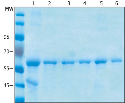
Western Blotting Analysis: 1 ug/mL from a representative lot detected ARF proteins in human lung tissue lysate.
Immunoprecipitation Analysis: A representative lot immunoprecipitated ARF proteins (Cavenagh, M.M., et. al. (1996). J Biol Chem. 271(36):21767-74).
ELISA Analysis: A representative lot detected pan-ARF in ELISA applications (Cavenagh, M.M., et. al. (1996). J Biol Chem. 271(36):21767-74).
Western Blotting Analysis: A representative lot detected ARF proteins in Western Blotting applications (Cavenagh, M.M., et. al. (1996). J Biol Chem. 271(36):21767-74; Zuezem, S., et. al. (1992). Proc Natl Acad Sci USA. 89(14):6619-23).
Electron Microscopy Analysis: A representative lot detected ARF proteins in Electron Microscopy applications (Zuezem, S., et. al. (1992). Proc Natl Acad Sci USA. 89(14):6619-23).
Dot Blot Analysis: A representative lot detected ARF proteins in Dot Blot applications (Cavenagh, M.M., et. al. (1996). J Biol Chem. 271(36):21767-74).
품질
Evaluated by Western Blotting in HeLa cell lysate.
Western Blotting Analysis: 1 ug/mL of this antibody detected pan-ARF in HeLa cell lysate.
Western Blotting Analysis: 1 ug/mL of this antibody detected pan-ARF in HeLa cell lysate.
표적 설명
~20 kDa observed. Uncharacterized bands may be observed in some lysate(s).
물리적 형태
Format: Purified
Protein G purified
Purified mouse monoclonal antibody IgG1 in buffer containing 0.1 M Tris-Glycine (pH 7.4), 150 mM NaCl with 0.05% sodium azide.
저장 및 안정성
Stable for 1 year at 2-8°C from date of receipt.
기타 정보
Concentration: Please refer to lot specific datasheet.
면책조항
Unless otherwise stated in our catalog or other company documentation accompanying the product(s), our products are intended for research use only and are not to be used for any other purpose, which includes but is not limited to, unauthorized commercial uses, in vitro diagnostic uses, ex vivo or in vivo therapeutic uses or any type of consumption or application to humans or animals.
적합한 제품을 찾을 수 없으신가요?
당사의 제품 선택기 도구.을(를) 시도해 보세요.
시험 성적서(COA)
제품의 로트/배치 번호를 입력하여 시험 성적서(COA)을 검색하십시오. 로트 및 배치 번호는 제품 라벨에 있는 ‘로트’ 또는 ‘배치’라는 용어 뒤에서 찾을 수 있습니다.
자사의 과학자팀은 생명 과학, 재료 과학, 화학 합성, 크로마토그래피, 분석 및 기타 많은 영역을 포함한 모든 과학 분야에 경험이 있습니다..
고객지원팀으로 연락바랍니다.