추천 제품
일반 설명
Neurogenic locus notch homolog protein 3 (UniProt Q9UM47; also known as Notch 3, Notch homolog 3) is encoded by the NOTCH3 (also known as CASIL, CADASIL, IMF2) gene (Gene ID 4854) in human. Notch 3 is a transmembrane receptor required for arterial differentiation and maturation of vascular smooth muscle cells in small arteries. Notch 3 is initially produced with a signal peptide (a.a. 1-39), the removal of which yields the 2282-amino acid Notch 3 (a.a. 40-2321), which undergoes a constitutive proteolytical cleavage, generating the extracellular fragment (Notch3-ECD; a.a. 40-1628) and the transmembrane/cytosolic (Notch3-TMIC; a.a. 1629-2321) fragment that remain associated at the cell surface to form a heterodimeric receptor. The ECD fragment contains all 34 EGF-like domains and the three Lin/Notch (LNR) repeats, while the extracellular portion of TMIC contains 5 ankyrin repeats. Mutations in the NOTCH3 gene have been identified as the underlying cause of cerebral autosomal dominant arteriopathy with subcortical infarcts and leukoencephalopathy (CADASIL). Pathogenic mutations predominantly affect cysteine residues within EGF-like repeats of Notch3-ECD, leading to Notch3-ECD multimerization and accumulation in the tunica media of vessel walls.
특이성
Clone 2G8 reacts with unprocessed Notch 3 and the extracellular (Notch 3-ECD; a.a. 40-1628) fragment, but not the transmembrane/cytosolic fragment (Notch 3-TMIC, also called Notch 3 extracellular truncation; a.a. 1629-2321) or the Notch 3 intracellular fragment (a.a. 1662-2321).
면역원
Epitope: Extracellular EGF-like domains 33-34.
KLH-conjugated linear peptide corresponding to a sequence from the EGF-like domains 33-34 region of human Notch 3 ECD.
애플리케이션
Anti-Notch 3 Antibody, ECD, clone 2G8 is an antibody against Notch 3 for use in Immunohistochemistry (Paraffin), Immunofluorescence, Western Blotting.
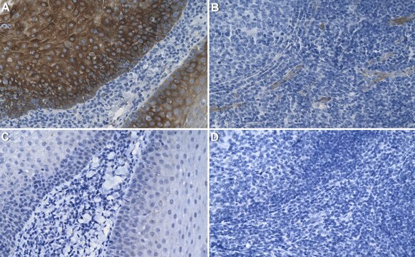
Immunohistochemistry Analysis: 20 µg/mL of this antibody from a representative lot detected Notch 3 extracellular domain (ECD) deposits in blood vessels of CADASIL patients brain tissue sections (Courtesy of Institute for Stroke and Dementia Research, Clinical center of the Ludwig-Maximilians University Munich, Germany).
Immunohistochemistry Analysis: A representative lot detected the aggregated Notch 3 extracellular domain (ECD) deposits in brain blood vessels of CADASIL patients by immunohistochemistry staining of paraformaldehyde-fixed, paraffin-embedded brain tissue sections (Kast, J., et al. (2014). Acta Neuropathol. Commun. 2:96).
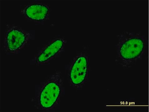
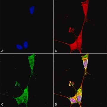
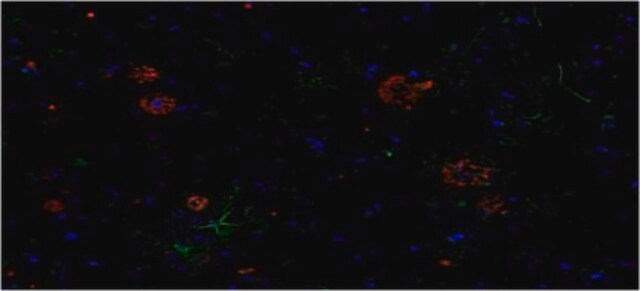
Immunofluorescence Analysis: A representative lot detected the aggregated Notch 3 extracellular domain (ECD) deposits in brain blood vessels of CADASIL patients by fluorescent immunohistochemistry staining of paraformaldehyde-fixed frozen brain tissue sections. The Notch3-EDC immunoreactivity was seen co-localized with that of LTBP-1, but not fibrillin-1 (Kast, J., et al. (2014). Acta Neuropathol. Commun. 2:96).
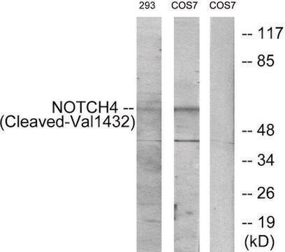
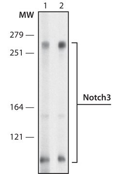
Western Blotting Analysis: A representative lot detected an elevated accumulation of aggregated Notch 3 extracellular domain (ECD) in brain blood vessel extracts from CADASIL patients when comparing to vessel extracts from non-diseased brains (Kast, J., et al. (2014). Acta Neuropathol. Commun. 2:96).
Research Category
Inflammation & Immunology
Inflammation & Immunology
Research Sub Category
Nervous System
Nervous System
품질
Identity Confirmation by Isotyping Test.
Isotyping Analysis: The identity of this monoclonal antibody is confirmed by isotyping test to be IgG2aκ.
Isotyping Analysis: The identity of this monoclonal antibody is confirmed by isotyping test to be IgG2aκ.
표적 설명
243.6 kDa (Pre-form), 239.5 kDa (mature, unprocessed), 167.13 kDa (Notch 3-ECD) calculated. ~250 kDa (Notch 3-ECD) reported (Kast, J., et al. (2014). Acta Neuropathol. Commun. 2:96).
물리적 형태
Format: Purified
Protein G Purified
Purified rat monoclonal IgG2aκ antibody in buffer containing 0.1 M Tris-Glycine (pH 7.4), 150 mM NaCl with 0.05% sodium azide.
저장 및 안정성
Stable for 1 year at 2-8°C from date of receipt.
기타 정보
Concentration: Please refer to lot specific datasheet.
면책조항
Unless otherwise stated in our catalog or other company documentation accompanying the product(s), our products are intended for research use only and are not to be used for any other purpose, which includes but is not limited to, unauthorized commercial uses, in vitro diagnostic uses, ex vivo or in vivo therapeutic uses or any type of consumption or application to humans or animals.
적합한 제품을 찾을 수 없으신가요?
당사의 제품 선택기 도구.을(를) 시도해 보세요.
Storage Class Code
12 - Non Combustible Liquids
WGK
WGK 1
Flash Point (°F)
Not applicable
Flash Point (°C)
Not applicable
시험 성적서(COA)
제품의 로트/배치 번호를 입력하여 시험 성적서(COA)을 검색하십시오. 로트 및 배치 번호는 제품 라벨에 있는 ‘로트’ 또는 ‘배치’라는 용어 뒤에서 찾을 수 있습니다.
자사의 과학자팀은 생명 과학, 재료 과학, 화학 합성, 크로마토그래피, 분석 및 기타 많은 영역을 포함한 모든 과학 분야에 경험이 있습니다..
고객지원팀으로 연락바랍니다.