
추천 제품


생물학적 소스
mouse
Quality Level
항체 형태
purified immunoglobulin
항체 생산 유형
primary antibodies
클론
5C9/C8, monoclonal
종 반응성
human, mouse
기술
ChIP: suitable
immunocytochemistry: suitable
immunohistochemistry: suitable (paraffin)
western blot: suitable
동형
IgG1κ
NCBI 수납 번호
UniProt 수납 번호
배송 상태
ambient
타겟 번역 후 변형
unmodified
유전자 정보
human ... TADA3(10474)
일반 설명
Transcriptional adapter 3 (UniProt O75528; also known as ADA3 homolog, ADA3-like protein, Alteration/deficiency in activation 3, hADA3, STAF54, Transcriptional adapter 3-like) is encoded by the TADA3 (also known as ADA3, TADA3L) gene (Gene ID 10474) in human. Alteration/deficiency in activation 3 (ADA3) is a component of several transcriptional co-activator and histone acetyltransferase (HAT) complexes and plays a critical role in in cell cycle regulation. Ada3 deletion in mice is embryonic lethal, and Ada3-deficient mouse embryonic fibroblasts (MEFs) exhiit a severe proliferation defect, dramatic changes in global histone acetylation, mitotic defects, as well as a delay in G2/M-to-G1 and G1-to-S transition. ADA3 also plays a role in genomic stability by controlling DNA repair checkpoints. ChIP-seq analsis reveals that ADA3 is significantly associated with human centromere regions across most chromosomes. In addition, ADA3 is found associated with CENP-B throughout all phases of the cell cycle, and CENP-B centromere binding decreased upon ADA3 knockdown. Wild-type human ADA3, but not CENP-B-binding deficient ADA3 mutant, prevented cell proliferation defects in MEFs following endogenous mouse ADA3 knockown. ADA3 overexpression and mislocalization correlates with poor prognosis in breast cancer patients.
특이성
Clone 5C9/C8 specifically detected Cre recombinase expression-induced ADA3 downregulation in Ada3FL/FL MEFs. Clone 5C9/C8 immunostained the nuclei of untransfected, but not ADA3 shRNA-transfected, 76N-TERT human mammary epithelial cells (Mohibi, S., et al. (2015). J. Biol. Chem. 290(47):28299-28310; Mohibi, S., et al. (2012). J. Biol. Chem. 287(35):29442-29456).
면역원
Full-length recombinant human ADA3.
애플리케이션
Anti-ADA3, clone 5C9/C8, Cat. No. MABE1057, is a highly specific mouse monoclonal antibody that targets TADA3 and has been tested in Chromatin Immunoprecipitation (ChIP), Immunocytochemistry, Immunohistochemistry (Paraffin), and Western Blotting.
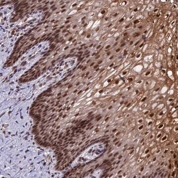
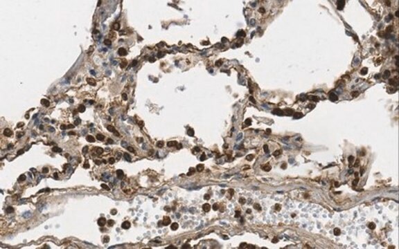
Immunohistochemistry Analysis: A representative lot detected nuclear ADA3 immunoreactivity in formalin-fixed, paraffin-embedded human breast carcinoma tissue section (Courtesy of Vimla Band, Ph.D., University of Nebraska USA).
Chromatin Immunoprecipitation (ChIP) Analysis: A representative lot detected ADA3 occupancy at the X chromosome centromere HOR region, the kinetochore assembly site also occupied by CENP-A and CENP-B, using TERT-immortalized human mammary epithelial cell 76N-TERT chromatin preparation. shRNA-mediated ADA3 knockdown led to a reduction in CENP-B, but not CENP-A, recruitment to the HOR region (Mohibi, S., et al. (2015). J. Biol. Chem. 290(47):28299-28310).
Immunocytochemistry Analysis: A representative lot detected ADA3 nuclear immunoreactivity in untransfected, but not ADA3 shRNA-transfected, TERT-immortalized human mammary epithelial 76N-TERT cells by fluorescent immunocytochemistry staining of 4% formaldehyde-fixed cells. Dual fluorescent staining revealed ADA3 and CENP-B nuclear colocalization (Mohibi, S., et al. (2015). J. Biol. Chem. 290(47):28299-28310).
Western Blotting Analysis: Representative lots detected both the endogenous mouse ADA3 (mADA3) and the exogenously expressed human ADA3 (hADA3) in Ada3FL/FL MEFs virally infected to express FLAG-tagged full-length or a.a. 111-432 hADA3 constructs. Cre recombinase expression downregulated only mADA3, but not hADA3 (Mohibi, S., et al. (2015). J. Biol. Chem. 290(47):28299-28310; Mohibi, S., et al. (2012). J. Biol. Chem. 287(35):29442-29456).
Western Blotting Analysis: A representative lot detected ADA3 expression levels among a panel of mouse tissues (Mohibi, S., et al. (2012). J. Biol. Chem. 287(35):29442-29456).
Research Category
Epigenetics & Nuclear Function
Epigenetics & Nuclear Function
품질
Evaluated by Western Blotting in MCF7 cell lysate.
Western Blotting Analysis: A 1:1,000 dilution of this antibody detected ADA3 in 10 µg of MCF-7 cell lysate.
Western Blotting Analysis: A 1:1,000 dilution of this antibody detected ADA3 in 10 µg of MCF-7 cell lysate.
표적 설명
~55 kDa observed. 48.90 kDa (human and mouse isoform 1) calculated. The larger-than-calculated band size is consistent with that reported in the literature (Mohibi, S., et al. (2015). J. Biol. Chem. 290(47):28299-28310). Uncharacterized bands may be observed in some lysate(s).
물리적 형태
Format: Purified
Protein G purified.
Purified mouse IgG1 in buffer containing 0.1 M Tris-Glycine (pH 7.4), 150 mM NaCl with 0.05% sodium azide
저장 및 안정성
Stable for 1 year at 2-8°C from date of receipt.
기타 정보
Concentration: Please refer to lot specific datasheet.
면책조항
Unless otherwise stated in our catalog or other company documentation accompanying the product(s), our products are intended for research use only and are not to be used for any other purpose, which includes but is not limited to, unauthorized commercial uses, in vitro diagnostic uses, ex vivo or in vivo therapeutic uses or any type of consumption or application to humans or animals.
적합한 제품을 찾을 수 없으신가요?
당사의 제품 선택기 도구.을(를) 시도해 보세요.
Storage Class Code
12 - Non Combustible Liquids
WGK
WGK 1
Flash Point (°F)
Not applicable
Flash Point (°C)
Not applicable
시험 성적서(COA)
제품의 로트/배치 번호를 입력하여 시험 성적서(COA)을 검색하십시오. 로트 및 배치 번호는 제품 라벨에 있는 ‘로트’ 또는 ‘배치’라는 용어 뒤에서 찾을 수 있습니다.
자사의 과학자팀은 생명 과학, 재료 과학, 화학 합성, 크로마토그래피, 분석 및 기타 많은 영역을 포함한 모든 과학 분야에 경험이 있습니다..
고객지원팀으로 연락바랍니다.