추천 제품
관련 카테고리
일반 설명
Programmed cell death 1 ligand 1 (UniProt: Q9EP73; also known as PD-L1, PDCD1 ligand 1, Programmed death ligand 1, B7 homolog 1, B7-H1, CD274) is encoded by the CD274 (also known as B7h1, Pdcd1l1, Pdcd1lg1, Pdl1) gene (Gene ID: 60533) in murine species. PD-1 and PD-1 ligands 1 and 2 (PD-L1 and PD-L2) are B7:CD28 family members that regulate T cell activation and peripheral tolerance. PD-L1 is initially produced with signal peptide (aa: 1-18) sequence, the removal of which yields the mature protein with a large extracellular (aa: 19-238) region that contains an Ig-like V-type domain (aa: 19-127) and an Ig-like C2-type domain (aa: 133-225), followed by a transmembrane domain (aa: 239-259) and a cytoplasmic tail (aa: 260-290). When engaged together with the TCR, the interaction of PD-1 with its ligands delivers an inhibitory signal to T cell proliferation and cytokine production. While PD-L1 is broadly expressed in hematopoietic and non-hematopoietic cells, PD-L2 expression is highly restricted to antigen presenting cells (APCs), including dendritic cells (DCs) and macrophages. The PD-1 pathway plays a key role in the progressive loss of effector T cell responses during chronic HIV infection. Tumor-associated PD-L1 is reported to facilitate apoptosis of activated T cells and stimulate IL-10 production in T cells to mediate immune suppression. These effects can be blocked by some monoclonal PD-L1 antibodies and T cell function can be restored. (Ref.: Chen, L., and Han, X (2015). J. Clin. Invest. 125(9): 3384-3391).
특이성
Clone 10F.9G2 specifically detects PD-L1 in murine EL4 lymphoma cells and targets the extracellular domain.
면역원
CHO-mPD-L1 transfectants.
애플리케이션
Detect PD-L1 using this rat monoclonal Anti-PD-L1 Antibody, clone 10F.9G2, Cat. No. MABC991. It has been tested in Flow Cytometry, Function Induction, Immunohistochemistry, and Inhibition studies.
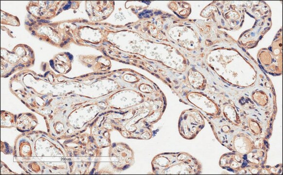
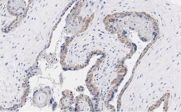
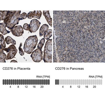
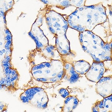
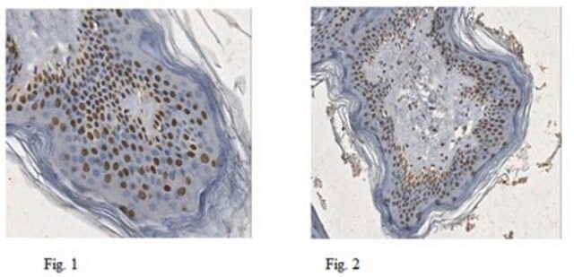
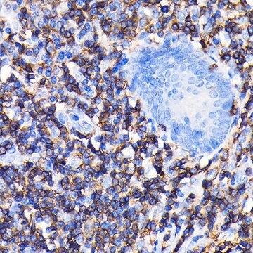
Immunohistochemistry Analysis: A representative lot detected PD-L1 in Immunohistochemistry applications (Grabie, N., et. al. (2007). Circulation. 116(18):2062-71).
Induces Function Analysis: Blockade with a representative lot of anti-PD-L1 combined with IL-2 enhanced antiviral CD8 T cell responses during chronic LCMV infection (West, E.E., et. al. (2013). J Clin Invest. 123(6):2604-15).
Inhibition Analysis: A representative lot blocked the action of PD-L1 (Grabie, N., et. al. (2007). Circulation. 116(18):2062-71;West, E.E., et. al. (2013). J Clin Invest. 123(6):2604-15;El Annan, J., et. al. (2010). Invest Ophthalmol Vis Sci. 51(7):3418-23).
품질
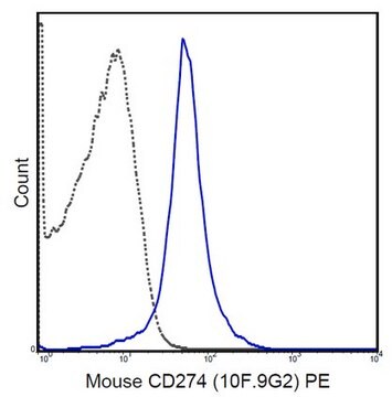
Evaluated by Flow Cytometry in EL4 cells.
Flow Cytometry Analysis: 1 µg/mL of this antibody detected PD-L1 in one million EL4 cells.
Flow Cytometry Analysis: 1 µg/mL of this antibody detected PD-L1 in one million EL4 cells.
표적 설명
32.78 kDa calculated.
결합
Replaces: MABF405
물리적 형태
Format: Purified
기타 정보
Concentration: Please refer to lot specific datasheet.
적합한 제품을 찾을 수 없으신가요?
당사의 제품 선택기 도구.을(를) 시도해 보세요.
Storage Class Code
12 - Non Combustible Liquids
WGK
WGK 2
Flash Point (°F)
Not applicable
Flash Point (°C)
Not applicable
시험 성적서(COA)
제품의 로트/배치 번호를 입력하여 시험 성적서(COA)을 검색하십시오. 로트 및 배치 번호는 제품 라벨에 있는 ‘로트’ 또는 ‘배치’라는 용어 뒤에서 찾을 수 있습니다.
자사의 과학자팀은 생명 과학, 재료 과학, 화학 합성, 크로마토그래피, 분석 및 기타 많은 영역을 포함한 모든 과학 분야에 경험이 있습니다..
고객지원팀으로 연락바랍니다.