추천 제품
생물학적 소스
mouse
Quality Level
항체 형태
purified immunoglobulin
항체 생산 유형
primary antibodies
클론
16H11.2, monoclonal
종 반응성
human
기술
immunohistochemistry: suitable
western blot: suitable
동형
IgG2bκ
NCBI 수납 번호
UniProt 수납 번호
배송 상태
wet ice
타겟 번역 후 변형
unmodified
유전자 정보
human ... LAMP3(27074)
일반 설명
Lysosyme-associated membrane proteins (LAMP) are transmembrane lysosomal glycoproteins, which share a common Gly-Tyr motif as a lysosomal targeting signal. While LAMP-1 and LAMP-2 contain type 1 transmembrane domains, LAMP-3 contains a transmembrane 4 superfamily domain, tetraspanin. FceRI-mediated stimulation of basophils causes LAMP-3 to be translocated from its intracellular localization to the plasma membrane. LAMP-3 thereby serves as a reliable marker for basophil activation. LAMP-3 expression is known to be reported in human bone marrow mast cells and in leukemic human mast cell line (HMC-1), among other cell lines. LAMP3 is shown to be regulated by hypoxia in a panel of tumor cells, via the unfolded protein response (UPR). UPR is a mechanism of adaptation to endoplasmic reticulum stress and is demonstrated to contribute to hypoxic adaptation in tumors through multiple mechanisms.
면역원
Recombinant protein corresponding to human LAMP-3.
애플리케이션
Detect LAMP-3 using this Anti-LAMP-3 Antibody, clone 16H11.2 validated for use in WB, IH.
Immunohistochemistry Analysis: A 1:200 dilution from a representative lot detected LAMP-3 in colorectal adenocarcinoma tissue.
Research Category
Cell Structure
Cell Structure
Research Sub Category
Apoptosis - Additional
Organelle & Cell Markers
Apoptosis - Additional
Organelle & Cell Markers
품질
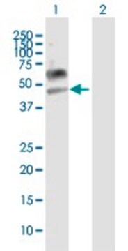
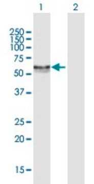
Evaluated by Western Blot in MCF-7 cell lysate.
Western Blot Analysis: 1 µg/mL of this antibody detected LAMP-3 in MCF-7 cell lysate.
Western Blot Analysis: 1 µg/mL of this antibody detected LAMP-3 in MCF-7 cell lysate.
표적 설명
~65 kDa observed.
The calculated molecular weight of this protein is 41 kDa, but has been observed at ~65-90 kDa due to glycosolation (de Saint-Vis, B., et al. (1998). Immunity. 9(3):325-336).
The calculated molecular weight of this protein is 41 kDa, but has been observed at ~65-90 kDa due to glycosolation (de Saint-Vis, B., et al. (1998). Immunity. 9(3):325-336).
물리적 형태
Format: Purified
Protein G Purified
Purified mouse monoclonal IgG2bκ in buffer containing 0.1 M Tris-Glycine (pH 7.4), 150 mM NaCl with 0.05% sodium azide.
저장 및 안정성
Stable for 1 year at 2-8°C from date of receipt.
분석 메모
Control
MCF-7 cell lysate
MCF-7 cell lysate
기타 정보
Concentration: Please refer to the Certificate of Analysis for the lot-specific concentration.
면책조항
Unless otherwise stated in our catalog or other company documentation accompanying the product(s), our products are intended for research use only and are not to be used for any other purpose, which includes but is not limited to, unauthorized commercial uses, in vitro diagnostic uses, ex vivo or in vivo therapeutic uses or any type of consumption or application to humans or animals.
적합한 제품을 찾을 수 없으신가요?
당사의 제품 선택기 도구.을(를) 시도해 보세요.
Storage Class Code
12 - Non Combustible Liquids
WGK
WGK 1
Flash Point (°F)
Not applicable
Flash Point (°C)
Not applicable
시험 성적서(COA)
제품의 로트/배치 번호를 입력하여 시험 성적서(COA)을 검색하십시오. 로트 및 배치 번호는 제품 라벨에 있는 ‘로트’ 또는 ‘배치’라는 용어 뒤에서 찾을 수 있습니다.
Junya Nishimura et al.
Esophagus : official journal of the Japan Esophageal Society, 16(4), 333-344 (2019-04-11)
Dendritic cells (DCs) are the most potent antigen-presenting cells to induce cytotoxic T lymphocytes in the tumor environment. After acquiring antigens, DCs undergo maturation and their expression of MHC and co-stimulation molecules are enhanced, along with lysosome-associated membrane glycoprotein 3
자사의 과학자팀은 생명 과학, 재료 과학, 화학 합성, 크로마토그래피, 분석 및 기타 많은 영역을 포함한 모든 과학 분야에 경험이 있습니다..
고객지원팀으로 연락바랍니다.
