추천 제품
생물학적 소스
hamster (Armenian)
항체 형태
purified immunoglobulin
항체 생산 유형
primary antibodies
클론
G4, monoclonal
종 반응성
mouse
포장
antibody small pack of 25 μg
기술
flow cytometry: suitable
NCBI 수납 번호
UniProt 수납 번호
타겟 번역 후 변형
unmodified
유전자 정보
mouse ... Pdcd1(18566)
관련 카테고리
일반 설명
Protein PD-1, mPD-1, CD279) is encoded by the PDCD1 (also known as PD1) gene (Gene ID: 18566) in murine species. PD-1 is a monomeric inhibitory cell surface receptor involved in the regulation of T-cell function during immunity and tolerance. PD-1 is synthesized with a signal peptide (aa 1-20), which is subsequently cleaved off. The mature form contains an extracellular domain (aa 21-169), a transmembrane domain (aa 170-190), and a cytoplasmic domain (aa 191-288). PD-1 also contains a single N-terminal immunoglobulin variable region (IgV) like domain (aa 31-139). PD-1 and its ligands, PD-L1 and PD-L2 play a key role in the maintenance of peripheral tolerance, a process by which the quiescence of autoreactive mature T cells is maintained. However, tumors and pathogens that cause chronic infections can exploit this pathway to escape T-cell mediated tumor-specific and pathogen-specific immunity. The effector functions of T-cells expressing PD-1 can be downregulated by PD-L1 or PD-L2 expressed by the tumor cells. PD-1 lacks SH2- or SH3-binding motifs on its cytoplasmic tail, but contains the N-terminal sequence VDYGEL that forms an immunoreceptor tyrosine-based inhibition motif (V/I/LxYxxL), which recruits SH2 domain-containing phosphatases. The cytoplasmic tail also contains the C-terminal sequence TEYATI, which forms an immunoreceptor tyrosine-based switch motif (TxYxxL). PD-1 ligation is reported to inhibit the activation of T-cell receptor proximal kinases, which results in attenuation of Lck-mediated phosphorylation of ZAP-70 and initiation of downstream events. PD-1 is also reported to impair the activation of the MEK-ERK MAP kinase pathway by inhibiting activation of PLC- 1 and Ras.
특이성
Clone G4 is an Armenian Hamster monoclonal antibody that specifically detects PD-1 in murine cells.
면역원
Murine PD-1Ig fusion protein.
애플리케이션
Affects Function Analysis: A representative lot detected PD-1 in Affects Funtion applications (Dronca, R.S., et. al. (2016). JCI Insight. 1(6); Hirano, F., et. al. (2005). Cancer Res. 65(3):1089-96; Overacre-Delgoffe, A.E., et. al. (2017). Cell. 169(6):1130; Tsushima, F., et. al. (2006). Blood. 110(1):180-5).
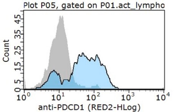
Flow Cytometry Analysis: A representative lot detected PD-1 in Flow Cytometry applications (Hirano, F., et. al. (2005). Cancer Res. 65(3):1089-96).
Flow Cytometry Analysis: A representative lot detected PD-1 in Flow Cytometry applications (Hirano, F., et. al. (2005). Cancer Res. 65(3):1089-96).
Detects Programmed cell death protein 1 using this armenian hamster monoclonal Anti-PD-1, clone G4, Cat. No. MABC1132, testted for use in Flow Cytometry and Affects Function.
품질
Evaluated by Flow Cytometry in EL4 T lymphoma cells.
Flow Cytometry Analysis: 1 µg of this antibody detected PD-1 in 1X10E6 EL4 T lymphoma cells.
Flow Cytometry Analysis: 1 µg of this antibody detected PD-1 in 1X10E6 EL4 T lymphoma cells.
표적 설명
~31.84 kDa calculated.
물리적 형태
Format: Purified
기타 정보
Concentration: Please refer to lot specific datasheet.
적합한 제품을 찾을 수 없으신가요?
당사의 제품 선택기 도구.을(를) 시도해 보세요.
시험 성적서(COA)
제품의 로트/배치 번호를 입력하여 시험 성적서(COA)을 검색하십시오. 로트 및 배치 번호는 제품 라벨에 있는 ‘로트’ 또는 ‘배치’라는 용어 뒤에서 찾을 수 있습니다.
Ti Wen et al.
Cancer immunology research, 10(2), 162-181 (2021-12-17)
Cytotoxic CD8+ T cells (CTL) are a crucial component of the immune system notable for their ability to eliminate rapidly proliferating malignant cells. However, the T-cell intrinsic factors required for human CTLs to accomplish highly efficient antitumor cytotoxicity are not
자사의 과학자팀은 생명 과학, 재료 과학, 화학 합성, 크로마토그래피, 분석 및 기타 많은 영역을 포함한 모든 과학 분야에 경험이 있습니다..
고객지원팀으로 연락바랍니다.