
MAB3392
Anti-Collagen Type III (COL3A1) Antibody
mouse monoclonal, 1E7-D7
동의어(들):
collagen, type III, alpha 1, collagen, fetal, Ehlers-Danlos syndrome type IV, autosomal dominant, alpha1 (III) collagen, collagen alpha-1(III) chain
로그인조직 및 계약 가격 보기
모든 사진(1)
About This Item
UNSPSC 코드:
12352203
eCl@ss:
32160702
NACRES:
NA.41
추천 제품


제품명
Anti-Collagen Type III Antibody, clone IE7-D7, clone 1E7-D7, from mouse
생물학적 소스
mouse
Quality Level
항체 형태
purified immunoglobulin
항체 생산 유형
primary antibodies
클론
1E7-D7, monoclonal
종 반응성
rat
종 반응성(상동성에 의해 예측)
human (based on 100% sequence homology)
기술
ELISA: suitable
immunohistochemistry: suitable
western blot: suitable
동형
IgG1κ
NCBI 수납 번호
UniProt 수납 번호
배송 상태
wet ice
타겟 번역 후 변형
unmodified
유전자 정보
human ... COL3A1(1281)
일반 설명
Type III collagen (also known as COL3A1), which adds structure and strength to connective tissues, is found in many places in the body, especially skin, lung, intestinal walls, and the walls of blood vessels. Collagen type III is initially produced as pro-collagen, a protein consisting of three pro-alpha1(III) chains that form the triple-stranded, rope-like molecule. After being synthesized, the pro-collagen molecule is modified by the cell. Enzymes modify the amino acids lysine and proline in the protein strands by adding chemical groups that are necessary for the strands to form a stable molecule and then later to crosslink to other molecules outside the cell. Other enzymes add sugars to the protein. The type III pro-collagen molecules are released from the cell and are processed by enzymes that clip small segments off either end of the molecules to form mature collagen. The mature collagen molecules assemble into fibrils. Cross-linking between molecules produces a very stable fibril, contributing to collagen′s tissue strengthening function.
특이성
This antibody detects collagen type III. There is no evidence for cross reactivity with Collagen Types I, V and VI or connective tissue proteins (Elastin, Fibronectin and Laminin) at suggested working concentrations.
면역원
Epitope: N-terminus
Human type III collagen (Werkmeister, J.A., et al. 1990).
애플리케이션
ELISA Analysis: A previous lot of this antibody was used in ELISA (Werkmeister, J.A., et al., 1991).
Western Blot Analysis: A previous lot of this antibody was used to detect collagen type III in western blot under non-reduced conditions (Werkmeister J.A., et al., 1988; Ramshaw, J.S., et al., 1988).
Some Collagen samples can be contaminated with other Collagen Types. When purified Collagen is used in an application the purity of the Collagen sample should be verified by SDS-page to minimize the risk of false positives.
Immunohistochemistry Analysis: A previous lot of this antibody was used to detect collagen type III in immunohistochemistry (Werkmeister J.A., et al., 1989; Werkmeister J.A., et al., 1989; Werkmeister J.A., et al., 1988).
Research Category
Cell Structure
Cell Structure
Research Sub Category
ECM Proteins
ECM Proteins
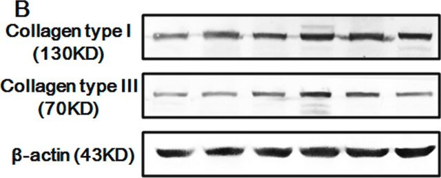
This Anti-Collagen Type III Antibody, clone IE7-D7 is validated for use in ELISA, WB, IH for the detection of Collagen Type III.
품질
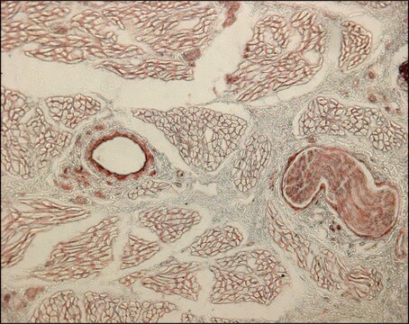
Evaluated by Immunohistochemistry in rat knee joint tissue.
Immunohistochemistry Analysis: A 1:600 dilution of this antibody detected Collagen Type III in rat knee joint tissue.
Immunohistochemistry Analysis: A 1:600 dilution of this antibody detected Collagen Type III in rat knee joint tissue.
표적 설명
138 kDa calculated
물리적 형태
Format: Purified
Protein G Purified
Purified mouse monoclonal IgG1κ in buffer containing 0.1 M Tris-Glycine (pH 7.4), 150 mM NaCl with 0.05% sodium azide.
저장 및 안정성
Stable for 1 year at 2-8°C from date of receipt.
분석 메모
Control
Rat knee joint tissue
Rat knee joint tissue
기타 정보
Concentration: Please refer to the Certificate of Analysis for the lot-specific concentration.
This clone displays a high affinity for human, dog, rat, kangaroo and porcine Type III Collagens.
면책조항
Unless otherwise stated in our catalog or other company documentation accompanying the product(s), our products are intended for research use only and are not to be used for any other purpose, which includes but is not limited to, unauthorized commercial uses, in vitro diagnostic uses, ex vivo or in vivo therapeutic uses or any type of consumption or application to humans or animals.
적합한 제품을 찾을 수 없으신가요?
당사의 제품 선택기 도구.을(를) 시도해 보세요.
Storage Class Code
12 - Non Combustible Liquids
WGK
WGK 1
Flash Point (°F)
Not applicable
Flash Point (°C)
Not applicable
시험 성적서(COA)
제품의 로트/배치 번호를 입력하여 시험 성적서(COA)을 검색하십시오. 로트 및 배치 번호는 제품 라벨에 있는 ‘로트’ 또는 ‘배치’라는 용어 뒤에서 찾을 수 있습니다.
Mingyu Cheng et al.
Tissue engineering. Part A, 16(5), 1479-1489 (2009-12-05)
Collagen-platelet (PL)-rich plasma composites have shown in vivo potential to stimulate anterior cruciate ligament (ACL) healing at early time points in large animal models. However, little is known about the cellular mechanisms by which the plasma component of these composites
Characterization of type I, III and V collagens in high-density cultured tenocytes by triple-immunofluorescence technique.
Gungormus, C; Kolankaya, D
Cytotechnology null
Sophie Cardin et al.
Circulation research, 100(3), 425-433 (2007-01-20)
Gene-expression changes in atrial fibrillation patients reflect both underlying heart-disease substrates and changes because of atrial fibrillation-induced atrial-tachycardia remodeling. These are difficult to separate in clinical investigations. This study assessed time-dependent mRNA expression-changes in canine models of atrial-tachycardia remodeling and
Lei Wang et al.
The Journal of biological chemistry, 289(2), 921-929 (2013-11-23)
Corneal stroma contains an extracellular matrix of orthogonal lamellae formed by parallel and equidistant fibrils with a homogeneous diameter of ~35 nm. This is indispensable for corneal transparency and mechanical functions. However, the mechanisms controlling corneal fibrillogenesis are incompletely understood
Michaela Leyh et al.
Stem cell research & therapy, 5(3), 77-77 (2014-06-12)
In the present study, we established a novel in vitro coculture model to evaluate the influence of osteoarthritis (OA) cartilage explants on the composition of newly produced matrix and chondrogenic differentiation of human bone marrow-derived mesenchymal stem cells (BMSCs) and
자사의 과학자팀은 생명 과학, 재료 과학, 화학 합성, 크로마토그래피, 분석 및 기타 많은 영역을 포함한 모든 과학 분야에 경험이 있습니다..
고객지원팀으로 연락바랍니다.