콘텐츠로 건너뛰기
Merck
모든 사진(1)

주요 문서

MAB1592-C

Sigma-Aldrich

Anti-Neurofilament NF-H Antibody, phosphorylated Antibody, clone NP1

clone NP1, from mouse

동의어(들):

Neurofilament heavy polypeptide, NF-H, 200 kDa neurofilament protein, Neurofilament triplet H protein

로그인조직 및 계약 가격 보기


About This Item

UNSPSC 코드:
12352203
eCl@ss:
32160702
NACRES:
NA.41

생물학적 소스

mouse

Quality Level

항체 형태

purified antibody

항체 생산 유형

primary antibodies

클론

NP1, monoclonal

종 반응성

canine, mouse, human, rat

종 반응성(상동성에 의해 예측)

chicken (based on 100% sequence homology), bovine (based on 100% sequence homology), porcine (based on 100% sequence homology)

포장

antibody small pack of 25 μg

기술

ELISA: suitable
immunohistochemistry: suitable (paraffin)
western blot: suitable

동형

IgG1κ

NCBI 수납 번호

UniProt 수납 번호

배송 상태

ambient

타겟 번역 후 변형

phosphorylation (not specified)

유전자 정보

bovine ... Nefh(528842)
chicken ... Nefh(417020)
dog ... Nefh(442940)
human ... NEFH(4744)
mouse ... Nefh(380684)
pig ... Nefh(100156492)
rat ... Nefh(24587)

일반 설명

Neurofilament heavy polypeptide (UniProt: P12036; also known as NF-H, 200 kDa neurofilament protein, Neurofilament triplet H protein) is encoded by the NEFH (also known as KIAA0845, NFH) gene (Gene ID: 4744) in human. Neurofilaments (~ 10 nm in diameter) are intermediate filaments that serve as major elements of the cytoskeleton supporting the axon cytoplasm. They are the most abundant fibrillar components of the axon, being on average 3-10 times more frequent than axonal microtubules. They are built from three intertwined protofibrils, which are themselves composed of two tetrameric protofilament complexes of monomeric proteins. The neurofilament triplet proteins (68/70, 160, and 200 kDa) occur in both the central and peripheral nervous system and are usually neuron specific. The 68/70 kDa NF-L protein can self-assemble into a filamentous structure, however the 160 kDa NF-M and 200 kDa NF-H proteins require the presence of the 68/70 kDa NF-L protein to co-assemble. Antibodies to NF-H are useful for identifying neuronal cells and their processes in tissue sections and in tissue culture. NF-H antibodies are helpful in the identification of neurofilament accumulations seen in many neurological diseases, such as Amyotrophic lateral sclerosis (ALS) and Alzheimer′s disease. The purified immunogen used to generate this antibody was the very heavily phosphorylated form of NF-H and this antibody binds only this heavily phosphorylated form. Similar, but far fewer phosphorylation sites are also found in NF-M. Hence, this antibody displays some reactivity with NF-M. Enzymatic phosphorylation of porcine and bovine neurofilaments is shown to result in loss of binding.

특이성

Clone NP1 detects heavily phosphorylated form of Neurofilament H. Displays weaker reactivity with phosphorylated Neurfilament -M.

면역원

Native Neurofilament H purified from pig spinal cord.

애플리케이션

Anti-Neurofilament NF-H, phosphorylated, clone NP1, Cat. No. MAB1592-C, is a highly specific mouse monoclonal antibody that targets Neurofilament heavy polypeptide and has been tested for use in ELISA, Immunohistochemistry (Paraffin), and Western Blotting.
Immunohistochemistry Analysis: A 1:50 dilution from a representative lot detected Neurofilament NF-H in human ovary tissue.

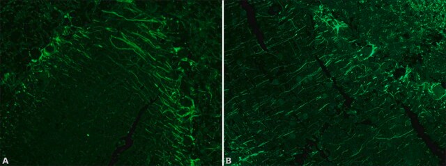
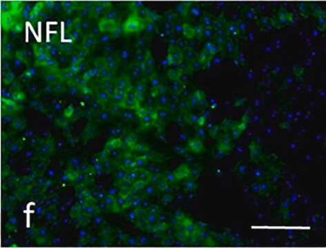
Immunohistochemistry Analysis: A representative lot detected Neurofilament NF-H in Immunohistochemistry applications (Benson, D.L., et. al. (1996). J Neurocytol. 25(3):181-96; Boylan, K., et. al. (2009). J Neurochem. 111(5):1182-91).

Western Blotting Analysis: A representative lot detected Neurofilament NF-H in Western Blotting applications (Benson, D.L., et. al. (1996). J Neurocytol. 25(3):181-96; Boylan, K., et. al. (2009). J Neurochem. 111(5):1182-91).

ELISA Analysis: A representative lot detected Neurofilament NF-H in ELISA applications (Boylan, K., et. al. (2009). J Neurochem. 111(5):1182-91; Toedebusch, C.M., et. al. (2017). J Vet Intem Med. 31(2):513-520; Mashita, T., et. al. (2015). J Vet Med Sci. 77(4):433-8).
Research Category
Neuroscience

품질

Evaluated by Immunohistochemistry in human cerebral cortex tissue.

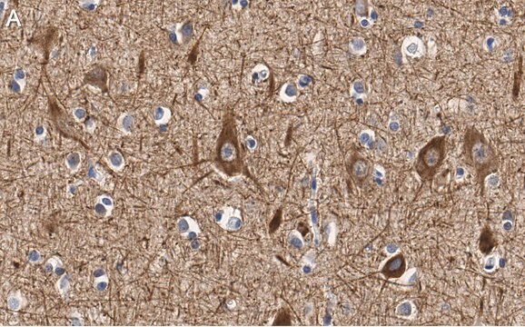
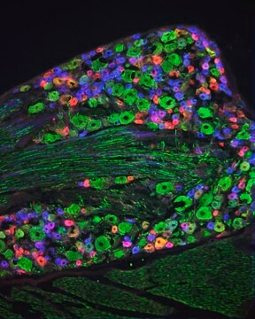
Immunohistochemistry Analysis: A 1:50 dilution of this antibody detected Neurofilament NF-H in human cerebral cortex tissue.

표적 설명

112.48 kDa calculated.

물리적 형태

Format: Purified
Protein G purified
Purified mouse monoclonal antibody IgG1 in PBS with 5 mM sodium azide and 50% glycerol.

저장 및 안정성

Stable for 1 year at -20°C from date of receipt.

Handling Recommendations: Upon receipt and prior to removing the cap, centrifuge the vial and gently mix the solution. Aliquot into microcentrifuge tubes and store at -20°C. Avoid repeated freeze/thaw cycles, which may damage IgG and affect product performance.

기타 정보

Concentration: Please refer to lot specific datasheet.

면책조항

Unless otherwise stated in our catalog or other company documentation accompanying the product(s), our products are intended for research use only and are not to be used for any other purpose, which includes but is not limited to, unauthorized commercial uses, in vitro diagnostic uses, ex vivo or in vivo therapeutic uses or any type of consumption or application to humans or animals.

적합한 제품을 찾을 수 없으신가요?  

당사의 제품 선택기 도구.을(를) 시도해 보세요.

Storage Class Code

12 - Non Combustible Liquids

WGK

WGK 2


시험 성적서(COA)

제품의 로트/배치 번호를 입력하여 시험 성적서(COA)을 검색하십시오. 로트 및 배치 번호는 제품 라벨에 있는 ‘로트’ 또는 ‘배치’라는 용어 뒤에서 찾을 수 있습니다.

이 제품을 이미 가지고 계십니까?

문서 라이브러리에서 최근에 구매한 제품에 대한 문서를 찾아보세요.

문서 라이브러리 방문

자사의 과학자팀은 생명 과학, 재료 과학, 화학 합성, 크로마토그래피, 분석 및 기타 많은 영역을 포함한 모든 과학 분야에 경험이 있습니다..

고객지원팀으로 연락바랍니다.