
추천 제품

생물학적 소스
mouse
항체 형태
purified immunoglobulin
항체 생산 유형
primary antibodies
클론
QBEnd/10, monoclonal
종 반응성
human, monkey
포장
antibody small pack of 25 μL
기술
electron microscopy: suitable
flow cytometry: suitable
immunofluorescence: suitable
immunohistochemistry: suitable (paraffin)
western blot: suitable
동형
IgG1λ
NCBI 수납 번호
UniProt 수납 번호
타겟 번역 후 변형
unmodified
유전자 정보
human ... CD34(947)
관련 카테고리
일반 설명
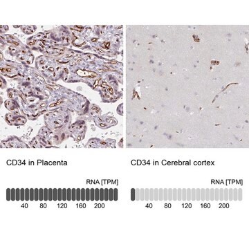
Hematopoietic progenitor cell antigen CD34 (UniProt: P28906; also known as CD34) is encoded by the CD34 gene (Gene ID: 947) in human. CD34 is a highly glycosylated single-pass type I membrane protein that is expressed on hematopoietic progenitor cells and small vessel endothelium of a variety of tissues. Under normal conditions, CD34+ expressing cells account for about 1 2% of the total bone marrow cells. It serves as an adhesion molecule that plays a role in early hematopoiesis by mediating the attachment of stem cells to the bone marrow extracellular matrix or directly to stromal cells. It is also reported to act as a scaffold for the attachment of lineage specific glycans, allowing stem cells to bind to lectins expressed by stromal cells or other marrow components. CD34 is synthesized with a signal peptide (aa 1-31) that is cleaved off in the mature form. The mature form has an extracellular domain (aa 32-290), a transmembrane domain (aa 291-311), and a cytoplasmic domain (aa 312-385). Two isoforms of CD34 have been described that are produced by alternative splicing.
특이성
Clone QBEnd/10 specifically detects CD34 in human and non-human primates.
면역원
Human placental endothelial membrane vesicles.
애플리케이션
Anti-CD34, clone QBEnd/10, Cat. No. CBL496-I, is a mouse monoclonal antibody that detects CD34 and has been tested for use in Electron Microscopy, Flow Cytometry, Immunofluorescence and Fluorescence Activated Cell Sorting (FACS), Immunohistochemistry (Paraffin), and Western Blotting.
Immunohistochemistry (Paraffin) Analysis: A 1:250 dilution from a representative lot detected CD34 in human brain tissue sections.
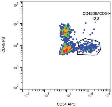
Fluorescence Activated Cell Sorting (FACS) Analysis: A representative lot was used to sort CD34+ cells from bone marrow. (de Bock, C.E., et. al. (2012). Leukemia. 26(5):918-26).
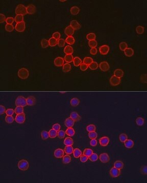
Immunofluorescence Analysis: A representative lot detected CD34 in Immunofluorescence applications (Miki, T., et. al. (2010). Mol Cancer Res. 8(5):665-76).
Electron Microscopy Analysis: A representative lot detected CD34 in Electron Microscopy applications (Fina, L., et. al. (1990). Blood. 75(12):2417-26).
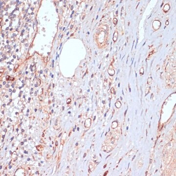
Immunohistochemistry Analysis: A representative lot detected CD34 in Immunohistochemistry applications (Engler, J.R., et. al. (2012). PLoS One. 7(8):e43339; Fina, L., et. al. (1990). Blood. 75(12):2417-26).
Flow Cytometry Analysis: A representative lot detected CD34 in Flow Cytometry applications (de Bock, C.E., et. al. (2012). Leukemia. 26(5):918-26; Fina, L., et. al. (1990). Blood. 75(12):2417-26).
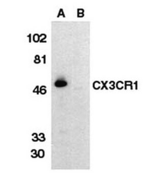
Western Blotting Analysis: A representative lot detected CD34 in Western Blotting applications (Fina, L., et. al. (1990). Blood. 75(12):2417-26).
Fluorescence Activated Cell Sorting (FACS) Analysis: A representative lot was used to sort CD34+ cells from bone marrow. (de Bock, C.E., et. al. (2012). Leukemia. 26(5):918-26).
Immunofluorescence Analysis: A representative lot detected CD34 in Immunofluorescence applications (Miki, T., et. al. (2010). Mol Cancer Res. 8(5):665-76).
Electron Microscopy Analysis: A representative lot detected CD34 in Electron Microscopy applications (Fina, L., et. al. (1990). Blood. 75(12):2417-26).
Immunohistochemistry Analysis: A representative lot detected CD34 in Immunohistochemistry applications (Engler, J.R., et. al. (2012). PLoS One. 7(8):e43339; Fina, L., et. al. (1990). Blood. 75(12):2417-26).
Flow Cytometry Analysis: A representative lot detected CD34 in Flow Cytometry applications (de Bock, C.E., et. al. (2012). Leukemia. 26(5):918-26; Fina, L., et. al. (1990). Blood. 75(12):2417-26).
Western Blotting Analysis: A representative lot detected CD34 in Western Blotting applications (Fina, L., et. al. (1990). Blood. 75(12):2417-26).
품질
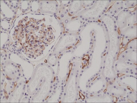
Evaluated by Immunohistochemistry (Paraffin) in human kidney tissue sections.
Immunohistochemistry (Paraffin) Analysis: A 1:250 dilution of this antibody detected CD34 in human kidney tissue sections.
Immunohistochemistry (Paraffin) Analysis: A 1:250 dilution of this antibody detected CD34 in human kidney tissue sections.
표적 설명
40.72 kDa Calculated. This antibody recognizes a heavily glycosylated transmembrane protein: gp 105-120 kDa
물리적 형태
Format: Purified
기타 정보
Concentration: Please refer to lot specific datasheet.
적합한 제품을 찾을 수 없으신가요?
당사의 제품 선택기 도구.을(를) 시도해 보세요.
시험 성적서(COA)
제품의 로트/배치 번호를 입력하여 시험 성적서(COA)을 검색하십시오. 로트 및 배치 번호는 제품 라벨에 있는 ‘로트’ 또는 ‘배치’라는 용어 뒤에서 찾을 수 있습니다.
자사의 과학자팀은 생명 과학, 재료 과학, 화학 합성, 크로마토그래피, 분석 및 기타 많은 영역을 포함한 모든 과학 분야에 경험이 있습니다..
고객지원팀으로 연락바랍니다.