추천 제품
생물학적 소스
mouse
Quality Level
항체 형태
purified immunoglobulin
항체 생산 유형
primary antibodies
클론
AE5, monoclonal
종 반응성
rabbit, bovine
기술
immunofluorescence: suitable
immunohistochemistry: suitable
western blot: suitable
동형
IgG1κ
NCBI 수납 번호
UniProt 수납 번호
배송 상태
wet ice
타겟 번역 후 변형
unmodified
유전자 정보
human ... KRT3(3850)
일반 설명
Cytokeratin 3, also known as 65kDa Cytokeratin or Keratin K3 is a specific keratin found primarily in the corneal epithelium and minorly in other epithelium like that of the esophagus. The corneal epithelium tissue forms the outermost layer of the cornea, which is the clear front covering of the eye. Keratins are divided into two primary types based upon their isoelectric mobility and size, acidic, or type 1:40-55kDa, and basic to neutral or type 2, 56-70kDa. Cytokeratin 3 is a type II cytokeratin. Type II cytokeratins form pairs with other cytokeratins. Cytokeratin 3 is expressed in the corneal epithelium with Cytokeratin 12. Together Cytokeratin 3 and Cytokeratin 12 form molecules known as intermediate filaments. These filaments assemble into strong networks that provide strength and resilience to the corneal epithelium. Mutations in Cytokeratin 3 have been associated with Meesmann′s Corneal Dystrophy a disease characterized by fragility of the anterior corneal epithelium.
면역원
Whole cells corresponding to Rabbit Keratin, type II cytosleletal 3.
애플리케이션
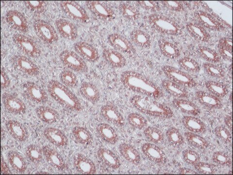
Immunohistochemistry Analysis: A 1:1000 dilution from a representative lot detected Keratin K3/K76 in bovine cornea epithelial cells.
Immunohistochemistry Analysis: A representative lot was used by an independent laboratory in Keratin 3 expressed epithelial cells on the surface of submerged and AL explants, indicating that they were derived from limbal progenitor cells via migration. (Kawakita, T., et al. (2005) Am J Pathol, 167:381-93.).
Western Blotting Analysis: A representative lot was used by an independent laboratory in Rabbit epithelial Keratin lysate. (Schermer, A, et al. (1986) J. Cell Biol., 103: 49-62).
Western Blotting Analysis: A representative lot was used by an independent laboratory in rabbit corneal tissue lysate. (Cooper, D., et al. (1986) J. Biol. Chem., 261: 4646-54).
Immunocytochemistry Analysis: A representative lot was used by an independent laboratory in human limbal stem cells cultured in medium supplemented with human EGF. (Sharifi, A.M., et al. (2010) Biocell. 34(1):53-55).
Immunofluorescence Analysis: A representative lot was used by an independent laboratory in rabbit corneal and limbal tissues, limbal explant, and epithelial outgrowth on human AM. (Wang D.Y., et al. (2003) Invest Ophthalmol Vis Sci. 44(11):4698-704).
Immunofluorescence Analysis: A representative lot was used by an independent laboratory inRabbit corneal epithelial Keratin cells. (Schermer, A, et al. (1986) J. Cell Biol., 103: 49-62).
Research Category
Cell Structure
Cell Structure
Research Sub Category
Cytokeratins
Cytokeratins
This Anti-Keratin K3/K76 Antibody, clone AE5 is validated for use in Western Blotting and Immunohistochemistry and Immunofluorescence for the detection of Keratin K3/K76.
품질
Evaluated by Western Blotting in Rabbit eye (cornea) cell lysate.
Western Blotting Analysis: 0.05 µg/mL of this antibody detected Keratin K3/K76 in 10 µg of Rabbit eye (cornea) cell lysate.
Western Blotting Analysis: 0.05 µg/mL of this antibody detected Keratin K3/K76 in 10 µg of Rabbit eye (cornea) cell lysate.
표적 설명
~60 kDa observed
결합
Replaces: CBL218
물리적 형태
Format: Purified
Protein G Purified
Purified mouse monoclonal IgG1κ in buffer containing 0.1 M Tris-Glycine (pH 7.4), 150 mM NaCl with 0.05% sodium azide.
저장 및 안정성
Stable for 1 year at 2-8°C from date of receipt.
기타 정보
Concentration: Please refer to lot specific datasheet.
면책조항
Unless otherwise stated in our catalog or other company documentation accompanying the product(s), our products are intended for research use only and are not to be used for any other purpose, which includes but is not limited to, unauthorized commercial uses, in vitro diagnostic uses, ex vivo or in vivo therapeutic uses or any type of consumption or application to humans or animals.
적합한 제품을 찾을 수 없으신가요?
당사의 제품 선택기 도구.을(를) 시도해 보세요.
Storage Class Code
12 - Non Combustible Liquids
WGK
WGK 1
Flash Point (°F)
Not applicable
Flash Point (°C)
Not applicable
시험 성적서(COA)
제품의 로트/배치 번호를 입력하여 시험 성적서(COA)을 검색하십시오. 로트 및 배치 번호는 제품 라벨에 있는 ‘로트’ 또는 ‘배치’라는 용어 뒤에서 찾을 수 있습니다.
Differentiation-related expression of a major 64K corneal keratin in vivo and in culture suggests limbal location of corneal epithelial stem cells.
Schermer, A; Galvin, S; Sun, TT
The Journal of cell biology null
Intrastromal invasion by limbal epithelial cells is mediated by epithelial-mesenchymal transition activated by air exposure.
Kawakita, T; Espana, EM; He, H; Li, W; Liu, CY; Tseng, SC
The American Journal of Pathology null
Monoclonal antibody analysis of bovine epithelial keratins. Specific pairs as defined by coexpression.
Cooper, D and Sun, T T
The Journal of Biological Chemistry, 261, 4646-4654 (1986)
Propagation and phenotypic preservation of rabbit limbal epithelial cells on amniotic membrane.
Wang, DY; Hsueh, YJ; Yang, VC; Chen, JK
Investigative Ophthalmology & Visual Science null
자사의 과학자팀은 생명 과학, 재료 과학, 화학 합성, 크로마토그래피, 분석 및 기타 많은 영역을 포함한 모든 과학 분야에 경험이 있습니다..
고객지원팀으로 연락바랍니다.

