
추천 제품


생물학적 소스
rabbit
Quality Level
항체 형태
affinity isolated antibody
항체 생산 유형
primary antibodies
클론
polyclonal
종 반응성
human, rat, bovine, mouse, chicken
기술
immunohistochemistry: suitable (paraffin)
immunoprecipitation (IP): suitable
western blot: suitable
동형
IgG
NCBI 수납 번호
UniProt 수납 번호
배송 상태
ambient
타겟 번역 후 변형
unmodified
유전자 정보
human ... ITPR1(3708)
rat ... Itpr1(25262)
일반 설명
Inositol 1,4,5-trisphosphate receptor type 1 (UniProt: P29994; also known as IP3 receptor isoform 1, IP-3-R, IP3R 1, InsP3R1, Type 1 inositol 1,4,5-trisphosphate receptor, Type 1 InsP3 receptor) is encoded by the Itpr1 (also known as Insp3r) gene (Gene ID: 25262) in rat. InsP3R1 is a multi-pass membrane protein located on endoplasmic reticulum (ER) membrane. Its highest levels are found in neurons, with very high expression in the Purkinje cells of the cerebellum. InsP3R1 contains a calcium channel in its C-terminal extremity. It has a large N-terminal cytoplasmic region that has the ligand-binding site in the N-terminus and modulatory sites in the middle portion immediately upstream of the channel region. InsP3R1 serves as an intracellular channel that mediates calcium release from the endoplasmic reticulum following stimulation by inositol 1,4,5-trisphosphate.It is involved in the regulation of epithelial secretion of electrolytes and fluid through the interaction with AHCYL1. It also plays a role in ER stress-induced apoptosis wherein cytoplasmic calcium released from the ER triggers apoptosis by the activation of CaM kinase II, eventually leading to the activation of downstream apoptosis pathways. InsP3R1 is phosphorylated by cAMP-dependent protein kinase (PKA), which increases the interaction with inositol 1,4,5-trisphosphate and reduces the interaction with AHCYL1. This prevents the ligand-induced opening of the calcium channels. Calcium appears to inhibit ligand binding to the receptor, most probably by interacting with a distinct calcium-binding protein which then inhibits the receptor. Reduced expression of InsP3R1 has been reported in cisplatin-resistant cancer cells and overexpression of InsP3R1 in resistant cells induces apoptosis and increases sensitivity to cisplatin. (Ref.: Tsunoda T., et al. (2005). Oncogene. 24(8):1396-402; Wang, Y., et al. (2001). Cir. Res. 88(2): 202-209).
특이성
This polyclonal antibody specifically react with IP3 Receptor 1 in multiple species, but does not recognize IP3R2 and IP3R3. It targets an epitope within 19 amino acids from the C-terminal region.
면역원
Epitope: C-terminus
KLH-conjugated linear peptide corresponding to 19 amino acids from the C-terminal region of rat Inositol 1,4,5-trisphosphate receptor type 1 (IP3R1).
애플리케이션
Detect Inositol 1,4,5-trisphosphate receptor type 1 using this rabbit polyclonal Anti-IP3R1, Cat. No. ABS2129 that has been tested for use in Immunohistochemistry (Paraffin), Immunoprecipitation, and Western Blotting. ,
Research Category
Signaling
Signaling
Western Blotting Analysis: A 1:1,000 dilution from a representative lot detected IP3R1 in 10 µg of rat and mouse brain microsomal preparation.
Western Blotting Analysis: A representative lot detected IP3R1 in bovine salivary gland, DT40 3KO cells stably expressing IP3R1 (Chandrasekhar, R., et. al. (2016). J Biol Chem. 291(10):4846-60).
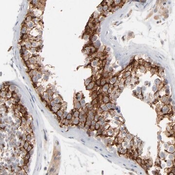
Immunohistochemistry Analysis: A 1:1,000 dilution from a representative lot detected IP3R1 in human brain and rat brain tissues.
Immunoprecipitation Analysis: A representative lot detected IP3R1 in bovine salivary gland (Chandrasekhar, R., et. al. (2016). J Biol Chem. 291(10):4846-60).
Western Blotting Analysis: A representative lot detected IP3R1 in bovine salivary gland, DT40 3KO cells stably expressing IP3R1 (Chandrasekhar, R., et. al. (2016). J Biol Chem. 291(10):4846-60).
Immunohistochemistry Analysis: A 1:1,000 dilution from a representative lot detected IP3R1 in human brain and rat brain tissues.
Immunoprecipitation Analysis: A representative lot detected IP3R1 in bovine salivary gland (Chandrasekhar, R., et. al. (2016). J Biol Chem. 291(10):4846-60).
품질
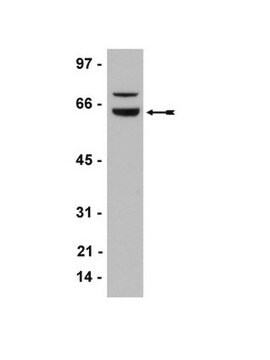
Evaluated by Western Blotting in rat brain microsomal preparation.
Western Blotting Analysis: A 1:1,000 dilution of this antibody detected IP3R1 in 10 µg of rat brain microsomal preparation.
Western Blotting Analysis: A 1:1,000 dilution of this antibody detected IP3R1 in 10 µg of rat brain microsomal preparation.
표적 설명
~300 kDa observed; 313.26 kDa calculated. Uncharacterized bands may be observed in some lysate(s).
물리적 형태
Affinity Purified
Format: Purified
Purified rabbit polyclonal antibody in PBS with 1% BSA and 0.02% sodium azide.
저장 및 안정성
Stable for 1 year at 2-8°C from date of receipt.
기타 정보
Concentration: Please refer to lot specific datasheet.
면책조항
Unless otherwise stated in our catalog or other company documentation accompanying the product(s), our products are intended for research use only and are not to be used for any other purpose, which includes but is not limited to, unauthorized commercial uses, in vitro diagnostic uses, ex vivo or in vivo therapeutic uses or any type of consumption or application to humans or animals.
적합한 제품을 찾을 수 없으신가요?
당사의 제품 선택기 도구.을(를) 시도해 보세요.
Storage Class Code
12 - Non Combustible Liquids
WGK
WGK 2
Flash Point (°F)
Not applicable
Flash Point (°C)
Not applicable
시험 성적서(COA)
제품의 로트/배치 번호를 입력하여 시험 성적서(COA)을 검색하십시오. 로트 및 배치 번호는 제품 라벨에 있는 ‘로트’ 또는 ‘배치’라는 용어 뒤에서 찾을 수 있습니다.
Yohannes A Mebratu et al.
Nature communications, 8(1), 803-803 (2017-10-08)
Bik reduces hyperplastic epithelial cells by releasing calcium from endoplasmic reticulum stores and causing apoptosis, but the detailed mechanisms are not known. Here we report that Bik dissociates the Bak/Bcl-2 complex to enrich for ER-associated Bak and interacts with the
자사의 과학자팀은 생명 과학, 재료 과학, 화학 합성, 크로마토그래피, 분석 및 기타 많은 영역을 포함한 모든 과학 분야에 경험이 있습니다..
고객지원팀으로 연락바랍니다.