
ABS1511
Anti-Neph1 Antibody, cytoplasmic domain
from rabbit, purified by affinity chromatography
동의어(들):
Kin of IRRE-like protein 1, Kin of irregular chiasm-like protein 1, Nephrin-like protein 1, Neph1, cytoplasmic domain
로그인조직 및 계약 가격 보기
모든 사진(1)
About This Item
UNSPSC 코드:
12352203
eCl@ss:
32160702
NACRES:
NA.41
추천 제품

생물학적 소스
rabbit
Quality Level
항체 형태
affinity isolated antibody
항체 생산 유형
primary antibodies
클론
polyclonal
정제법
affinity chromatography
종 반응성
mouse, rat, human
기술
electron microscopy: suitable
immunocytochemistry: suitable
immunofluorescence: suitable
immunoprecipitation (IP): suitable
western blot: suitable
NCBI 수납 번호
UniProt 수납 번호
배송 상태
wet ice
타겟 번역 후 변형
unmodified
유전자 정보
human ... KIRREL1(55243)
일반 설명
Kin of IRRE-like protein 1 (UniProt Q80W68; also known as Nephrin-like protein 1, Kin of IRRE like 1, Kin of irregular chiasm-like protein 1) is encoded by the Kirrel (also known as 6720469N11Rik, Kirrel1, Neph1) gene (Gene ID 170643) in murine species. Podocytes are specialized epithelial cells that are critical components of the glomerular filtration barrier. Glomerular injury, such as that caused by renal ischemia, is often characterized by the effacement of podocytes, loss of slit diaphragms, and proteinuria. The podocyte proteins Neph1 and nephrin are critical for maintaining the structural integrity of slit diaphragm and therefore the filtration function of the glomerulus by organizing a signaling complex at the podocyte cell membrane. Cytoplasmic domain of Neph1 plays a key role in actin cytoskeleton remodeling events that are directly associated with Fyn-mediated Neph1 pTyr637/638 phosphorylation that leads to the recruitment of adapter proteins and signaling molecules, such as Grb2, Csk tyrosine kinase, and ZO-1. Phosphorylation-induced association between Neph1 and ZO-1 plays a key role in defining the tight junction formation in podocytes.
특이성
Expected to react with murine spliced isoforms 1&2, as well as all three human spliced isoforms.
면역원
Epitope: cytoplasmic domain
His-tagged recombinant protein corresponding to the cytoplasmic domain of mouse Neph1.
애플리케이션
Detect Neph1 using this rabbit Polyclonal Anti-Neph1 Antibody, cytoplasmic domain, Cat. No. ABS1511, validated for use in Western Blotting, Immunoprecipitation, Immunofluorescence, Electron Microscopy and Immunocytochemistry.
Research Category
Signaling
Signaling
Research Sub Category
Signaling Neuroscience
Signaling Neuroscience
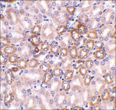
Western Blotting Analysis: A representative lot detected endogenous rat glomeruli Neph1 and exogenously expressed mouse Neph1 in COS-7 cells (Barletta, G.M., et al. (2003). J Biol Chem. 278(21):19266-19271).
Western Blotting Analysis: A representative lot detected ischemia-induced Neph1 membrane-to-cytosol translocation in human podocytes (Wagner, M.C., et al. (2008). J Biol Chem. 283(51):35579-35589).
Western Blotting Analysis: A representative lot detected Neph1 in mouse glomeruli & cultured human podocytes (Arif, E., et al. (2011). Mol Cell Biol. 31(10):2134-2150).
Immunoprecipitation Analysis: A representative lot immunoprecipitated Neph1 from rat glomerular and human podocyte cell lysates (Arif, E., et al. (2011). Mol Cell Biol. 31(10):2134-2150).
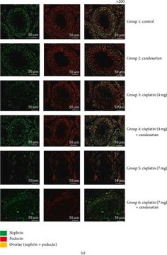
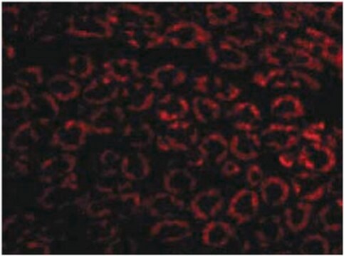
Immunofluorescence Analysis: A representative lot detected Neph1 using both paraffin-embedded and frozen rat kidney sections (Arif, E., et al. (2011). Mol Cell Biol. 31(10):2134-2150; Barletta, G.M., et al. (2003). J Biol Chem. 278(21):19266-19271).
Electron Microscopy Analysis: A representative lot detected Neph1 in frozen rat kidney sections (Barletta, G.M., et al. (2003). J Biol Chem. 278(21):19266-19271).
Immunocytochemistry Analysis: A representative lot detected Neph1 in cultured human podocytes (Arif, E., et al. (2014). J Biol Chem. 289(14):9502-9518; Arif, E., et al. (2011). Mol Cell Biol. 31(10):2134-2150; Wagner, M.C., et al. (2008). J Biol Chem. 283(51):35579-35589).
Western Blotting Analysis: A representative lot detected ischemia-induced Neph1 membrane-to-cytosol translocation in human podocytes (Wagner, M.C., et al. (2008). J Biol Chem. 283(51):35579-35589).
Western Blotting Analysis: A representative lot detected Neph1 in mouse glomeruli & cultured human podocytes (Arif, E., et al. (2011). Mol Cell Biol. 31(10):2134-2150).
Immunoprecipitation Analysis: A representative lot immunoprecipitated Neph1 from rat glomerular and human podocyte cell lysates (Arif, E., et al. (2011). Mol Cell Biol. 31(10):2134-2150).
Immunofluorescence Analysis: A representative lot detected Neph1 using both paraffin-embedded and frozen rat kidney sections (Arif, E., et al. (2011). Mol Cell Biol. 31(10):2134-2150; Barletta, G.M., et al. (2003). J Biol Chem. 278(21):19266-19271).
Electron Microscopy Analysis: A representative lot detected Neph1 in frozen rat kidney sections (Barletta, G.M., et al. (2003). J Biol Chem. 278(21):19266-19271).
Immunocytochemistry Analysis: A representative lot detected Neph1 in cultured human podocytes (Arif, E., et al. (2014). J Biol Chem. 289(14):9502-9518; Arif, E., et al. (2011). Mol Cell Biol. 31(10):2134-2150; Wagner, M.C., et al. (2008). J Biol Chem. 283(51):35579-35589).
품질
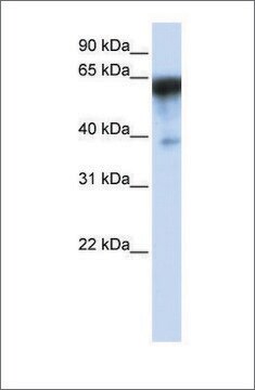
Evaluated by Western Blotting in rat kidney tissue lysate.
Western Blotting Analysis: A 1:1,000 dilution of this antibody detected Neph1 in rat kidney tissue lysate.
Western Blotting Analysis: A 1:1,000 dilution of this antibody detected Neph1 in rat kidney tissue lysate.
표적 설명
~110 kDa observed. Target band size appears larger than the calculated molecular weight (87.2 kDa/murine isoform 1, 70.0 kDa/murine isoform 2, 72.4-85.0 kDa/human isoform 1-3) due to posttranslational modifications (glycosylation & phosphorylation). An uncharacterized band ~25 kDa can also be seen in some samples.
물리적 형태
Affinity purified
Purified rabbit polyclonal antibody in buffer containing PBS/glycine/5% BSA, .02% Sodium Azide and 30% glycerol.
저장 및 안정성
Stable for 1 year at -20°C from date of receipt.
Handling Recommendations: Upon receipt and prior to removing the cap, centrifuge the vial and gently mix the solution. Aliquot into microcentrifuge tubes and store at -20°C. Avoid repeated freeze/thaw cycles, which may damage IgG and affect product performance.
Note: Variability in freezer temperatures below -20°C may cause glycerol containing solutions to become frozen during storage.
기타 정보
Concentration: Please refer to lot specific datasheet.
면책조항
Unless otherwise stated in our catalog or other company documentation accompanying the product(s), our products are intended for research use only and are not to be used for any other purpose, which includes but is not limited to, unauthorized commercial uses, in vitro diagnostic uses, ex vivo or in vivo therapeutic uses or any type of consumption or application to humans or animals.
적합한 제품을 찾을 수 없으신가요?
당사의 제품 선택기 도구.을(를) 시도해 보세요.
Storage Class Code
10 - Combustible liquids
WGK
WGK 3
시험 성적서(COA)
제품의 로트/배치 번호를 입력하여 시험 성적서(COA)을 검색하십시오. 로트 및 배치 번호는 제품 라벨에 있는 ‘로트’ 또는 ‘배치’라는 용어 뒤에서 찾을 수 있습니다.
Nephrin localizes to the slit pore of the glomerular epithelial cell.
Holzman, LB; St John, PL; Kovari, IA; Verma, R; Holthofer, H; Abrahamson, DR
Kidney International null
Mark C Wagner et al.
The Journal of biological chemistry, 283(51), 35579-35589 (2008-10-17)
Glomerular injury is often characterized by the effacement of podocytes, loss of slit diaphragms, and proteinuria. Renal ischemia or the loss of blood flow to the kidneys has been widely associated with tubular and endothelial injury but rarely has been
E Arif et al.
Molecular and cellular biology, 31(10), 2134-2150 (2011-03-16)
The podocyte proteins Neph1 and nephrin organize a signaling complex at the podocyte cell membrane that forms the structural framework for a functional glomerular filtration barrier. Mechanisms regulating the movement of these proteins to and from the membrane are currently
Ehtesham Arif et al.
The Journal of biological chemistry, 289(14), 9502-9518 (2014-02-21)
Podocytes are specialized epithelial cells that are critical components of the glomerular filtration barrier, and their dysfunction leads to proteinuria and renal failure. Therefore, preserving podocyte function is therapeutically significant. In this study, we identified Neph1 signaling as a therapeutic
Gina-Marie Barletta et al.
The Journal of biological chemistry, 278(21), 19266-19271 (2003-03-21)
Glomerular visceral epithelial cells (podocytes) appear to play a central role in maintaining the selective filtration barrier of the renal glomerulus. While the immunoglobulin superfamily member Nephrin was proposed to act as a cell adhesion molecule at the podocyte intercellular
자사의 과학자팀은 생명 과학, 재료 과학, 화학 합성, 크로마토그래피, 분석 및 기타 많은 영역을 포함한 모든 과학 분야에 경험이 있습니다..
고객지원팀으로 연락바랍니다.