추천 제품
생물학적 소스
rabbit
Quality Level
항체 형태
purified antibody
항체 생산 유형
primary antibodies
클론
polyclonal
종 반응성
mouse, human
포장
antibody small pack of 25 μg
기술
ChIP: suitable (ChIP-chip)
immunofluorescence: suitable
western blot: suitable
동형
IgG
NCBI 수납 번호
타겟 번역 후 변형
unmodified
유전자 정보
human ... MEIS1(4211)
mouse ... Meis1(17268)
일반 설명
Homeobox protein Meis1 (UniProt: Q60954-1; P97367-2 and P97367-3; also known as Myeloid ecotropic viral integration site 1 (Meis1) and Meis1-related protein 1) is encoded by the Meis1 gene (Gene ID: 17268). Meis proteins act as transcription factors that are orthologous to the Drosophila homothorax (Hth) protein. Meis protein contain a TALE (three-amino-acid loop extension) sub-class of the homeodomain that binds to DNA. Meis protein directly bind to Pbx proteins and the Meis/Pbx protein complex binds to DNA through respective Meis- and Pbx-consensus binding sites to regulate transcription. The Meis/Pbx complex plays important roles during development of multiple organs. In humans and mice, three homologues Meis1, Meis2 and Meis3 have been identified. Meis1 is expressed at high levels in the lung and at lower levels in the heart and brain. On the other hand, Meis 2 displays spatially restricted expression patterns in the developing nervous system, limbs, face, and in various viscera. In adult, it is mainly expressed in the brain and female genital tract, with a different distribution of the alternative splice forms in these organs. Pancreatic beta cells express both Meis1 and 2. Both Meis1 and 2 are expressed at high levels in all stages of embryonic development of mice (day 7 to 17). Deficiency of Meis1 or Meis 2 can lead to embryonic lethality. (Ref.: Machon, O et al. (2015). BMC Dev. Biol. 515:40).
특이성
This rabbit polyclonal antibody detects Meis1 in human and murine cells. It targets an epitope within the C-terminal region.
면역원
Epitope: C-terminus
KLH-conjugated linear peptide corresponding 14 amino acids from the C-terminal region of Meis1.
애플리케이션
Anti-Meis1, Cat. No. ABE2864, is a highly specific rabbit polyclonal antibody that targets Homeobox protein Meis1 has been tested in Chromatin Immunoprecipitation (ChIP), Immunofluorescence, and Western Blotting.
Research Category
Epigenetics & Nuclear Function
Epigenetics & Nuclear Function
Western Blotting Analysis: 1 µg/mL of this antibody detected Meis1 in 10 µg of K562 cell lysate.
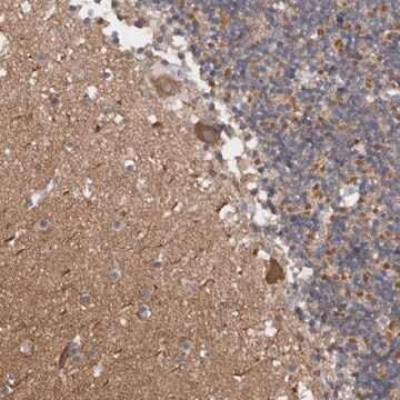
Immunofluorescence Analysis: A representative lot detected Meis1 in adjacent stage (St) 41 sagittal hindbrain and eye sections (Mercader, N., et. al. (2005). Development. 132(18):4131-42).
Western Blotting Analysis: A 1:800 dilution from a representative lot detected Meis1 in HEK293T cells transfectd with Meis1a (Courtesy of Dr.Valeria Azcoitia and Dr. Miguel Torres).
Chromatin Immunoprecipitation (ChIP) Analysis: A representative lot detected Meis1 in Chromatin Immunoprecipitation applications (Penkov, D., et. al. (2013). Cell Rep. 3(4):1321-33).
Western Blotting Analysis: A representative lot detected Meis1 in proximal, distal, and RA-treated distal blastemas (Mercader, N., et. al. (2005). Development. 132(18):4131-42).
품질
Evaluated by Western Blotting in HeLa cell lysate.
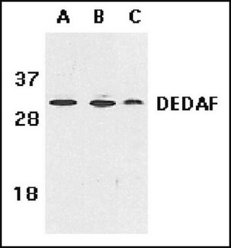
Western Blotting Analysis: 1 µg/mL of this antibody detected Meis1 in 10 µg of HeLa cell lysate.
Western Blotting Analysis: 1 µg/mL of this antibody detected Meis1 in 10 µg of HeLa cell lysate.
표적 설명
~52 kDa observed; 43.0 kDa (Meis1) calculated. Uncharacterized bands may be observed in some lysate(s).
물리적 형태
Format: Purified
Protein A purified
Purified rabbit polyclonal antibody in PBS with 0.05% sodium azide and 50% glycerol.
저장 및 안정성
Stable for 1 year at -20°C from date of receipt.
Handling Recommendations: Upon receipt and prior to removing the cap, centrifuge the vial and gently mix the solution. Aliquot into microcentrifuge tubes and store at -20°C. Avoid repeated freeze/thaw cycles, which may damage IgG and affect product performance.
Handling Recommendations: Upon receipt and prior to removing the cap, centrifuge the vial and gently mix the solution. Aliquot into microcentrifuge tubes and store at -20°C. Avoid repeated freeze/thaw cycles, which may damage IgG and affect product performance.
기타 정보
Concentration: Please refer to lot specific datasheet.
면책조항
Unless otherwise stated in our catalog or other company documentation accompanying the product(s), our products are intended for research use only and are not to be used for any other purpose, which includes but is not limited to, unauthorized commercial uses, in vitro diagnostic uses, ex vivo or in vivo therapeutic uses or any type of consumption or application to humans or animals.
적합한 제품을 찾을 수 없으신가요?
당사의 제품 선택기 도구.을(를) 시도해 보세요.
Storage Class Code
12 - Non Combustible Liquids
WGK
WGK 2
Flash Point (°F)
Not applicable
Flash Point (°C)
Not applicable
시험 성적서(COA)
제품의 로트/배치 번호를 입력하여 시험 성적서(COA)을 검색하십시오. 로트 및 배치 번호는 제품 라벨에 있는 ‘로트’ 또는 ‘배치’라는 용어 뒤에서 찾을 수 있습니다.
자사의 과학자팀은 생명 과학, 재료 과학, 화학 합성, 크로마토그래피, 분석 및 기타 많은 영역을 포함한 모든 과학 분야에 경험이 있습니다..
고객지원팀으로 연락바랍니다.