추천 제품
생물학적 소스
rabbit
Quality Level
항체 형태
purified antibody
항체 생산 유형
primary antibodies
분자량
calculated mol wt 15.4 kDa
observed mol wt ~17 kDa
정제법
affinity chromatography
종 반응성
human, mouse
포장
antibody small pack of 100 μL
기술
ChIP: suitable
dot blot: suitable
immunofluorescence: suitable
western blot: suitable
동형
IgG
에피토프 서열
N-terminal half
단백질 ID 수납 번호
UniProt 수납 번호
저장 온도
2-8°C
특이성
This rabbit polyclonal antibody specifically detects Histone H3 with butyrlated lysine 27.
면역원
KLH-conjugated linear peptide corresponding to 10 amino acids surrounding butyrlated lysine 27 of human Histone H3.
애플리케이션
Quality Control Testing
Evaluated by Western Blotting in lysates from HCT-116 cells treated with 5 mM Sodium butyrate.
Western Blotting Analysis: A 1:1,000 dilution of this antibody detected Histone H3 butyrlated on lysine 27 in lysate from HCT-116 cells treated with 5 mM Sodium butyrate.
Tested Applications
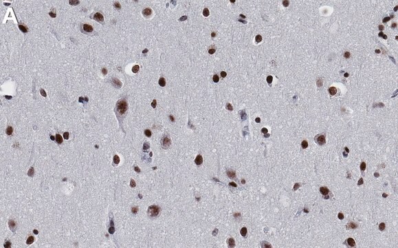
Immunofluorescence Analysis: A 1:500 dilution from a representative lot detected Histone H3K27butyrl in Mouse cecal sections (Data courtesy of Dr. Leah Gates, Dave Allis Lab @ Rockefeller University, New York).
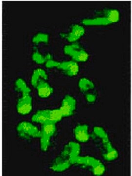
Chromatin Immunoprecipitation (ChIP) Analysis: 10 µg from a representative lot detected Histone H3K27butyrl in HCT-116 cells (Data courtesy of Dr. Leah Gates, Dave Allis Lab @ Rockefeller University, New York).
Dot Blot: A 1:2,000 dilution from a representative lot detected Histone H3K27butyrl peptide (Data courtesy of Dr. Leah Gates, Dave Allis Lab @ Rockefeller University, New York).
Note: Actual optimal working dilutions must be determined by end user as specimens, and experimental conditions may vary with the end user.
표적 설명
Histone H3 (UniProt: P68431; also known as Histone H3/a, Histone H3/b, Histone H3/c, Histone H3/d, Histone H3/f, Histone H3/h, Histone H3/I, Histone H3/j, Histone H3/k, Histone H3/l) is encoded by the HIST1H3A (also known as H3FA, HIST1H3B, H3FL, HIST1H3C, H3FC, HIST1H3D, H3FB, HISTH3F, H3FD, HIST1H3F, H3FI, HIST1H3G, H3FH, HIST1H3H, H3FK, HIST1H3I, H3FF, HIST1H3J, H3FJ) gene (Gene ID: 8350, 8351, 8352, 8354, 8355, 8356, 8357, 8358, 8968) in human. Histones and DNA assemble to form nucleosomes and each nucleosome organizes a stretch of DNA wrapped around a histone octamer that is composed of a central (H3-H4)2 tetramer, flanked by two H2A-H2B dimers. A large number of variants of Histone H3 have been described in mammals. The two main variants, H3.1 and H3.3, show different genomic localization patterns in animals. They possess distinct histone chaperones, CAF-1 and HIRA, which play important role in mediating DNA-synthesis-dependent and -independent nucleosome assembly. Histone H3.1 serves as the canonical histone, which is incorporated during DNA replication, whereas H3.3 acts as the replacement histone that can be incorporated outside of S-phase during chromatin-disrupting processes like transcription. Histone H3 features a main globular domain and a long N-terminal tail, which protrudes from the globular nucleosome core and can undergo several different types of epigenetic modifications that influence cellular processes. The amino-terminal tails of histone proteins are subject to several posttranslational modifications, including acetylation, methylation, and phosphorylation, which recruit downstream regulatory factors, influence chromatin structure, and are critical determinants of transcription. These modifications on histones, known as histone acyl marks, are involved in transcription regulation. However, the exact role of other modifications, such as propionylation and butyrlation are still under investigation. These modifications are detected at comparable levels to acetylation on selected residues in histone H3. Butyrlation of lysine 9 in histone H3 that occurs in cecal epithelial cells is shown to shown to decrease upon antibiotic treatment. This diminution can be rescued by treatment with exogenous tributyrin. (Ref.: Scott, WA., and Campos, EI. (2020). Front. Cell Dev. Biol. 8; 701; Allis CD., and Jenuwein, T. (20160. Nat. Rev. Genet. 17(8); 487-500).
물리적 형태
Purified rabbit polyclonal antibody in buffer containing 0.1 M Tris-Glycine (pH 7.4), 150 mM NaCl with 0.05% sodium azide.
재구성
0.5 mg/mL. Please refer to guidance on suggested starting dilutions and/or titers per application and sample type.
저장 및 안정성
Recommended storage: +2°C to +8°C.
기타 정보
Concentration: Please refer to the Certificate of Analysis for the lot-specific concentration.
면책조항
Unless otherwise stated in our catalog or other company documentation accompanying the product(s), our products are intended for research use only and are not to be used for any other purpose, which includes but is not limited to, unauthorized commercial uses, in vitro diagnostic uses, ex vivo or in vivo therapeutic uses or any type of consumption or application to humans or animals.
적합한 제품을 찾을 수 없으신가요?
당사의 제품 선택기 도구.을(를) 시도해 보세요.
Storage Class Code
12 - Non Combustible Liquids
WGK
WGK 1
Flash Point (°F)
Not applicable
Flash Point (°C)
Not applicable
시험 성적서(COA)
제품의 로트/배치 번호를 입력하여 시험 성적서(COA)을 검색하십시오. 로트 및 배치 번호는 제품 라벨에 있는 ‘로트’ 또는 ‘배치’라는 용어 뒤에서 찾을 수 있습니다.
자사의 과학자팀은 생명 과학, 재료 과학, 화학 합성, 크로마토그래피, 분석 및 기타 많은 영역을 포함한 모든 과학 분야에 경험이 있습니다..
고객지원팀으로 연락바랍니다.