추천 제품
생물학적 소스
rabbit
Quality Level
항체 형태
affinity purified immunoglobulin
항체 생산 유형
primary antibodies
클론
polyclonal
정제법
affinity chromatography
종 반응성
mouse, human
제조업체/상표
Chemicon®
기술
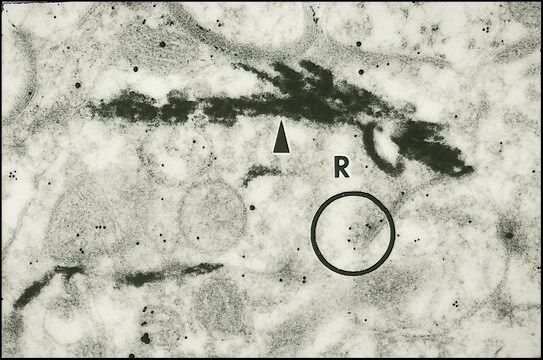
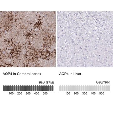
immunohistochemistry: suitable
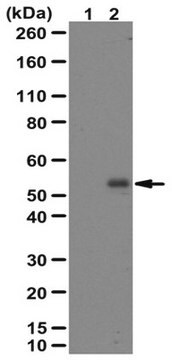
western blot: suitable
NCBI 수납 번호
UniProt 수납 번호
배송 상태
dry ice
타겟 번역 후 변형
unmodified
유전자 정보
human ... DLX2(1746)
특이성
Recognizes Dlx 2 (Distalless (DII) family), a homeodomain transcription factor.
면역원
Synthetic peptide, amino acids 24-39 from human Dlx 2.
애플리케이션
Anti-Dlx2 Antibody is an antibody against Dlx2 for use in WB, IH.
Research Category
Neuroscience
Neuroscience
Research Sub Category
Developmental Neuroscience
Neuronal & Glial Markers
Developmental Neuroscience
Neuronal & Glial Markers
Western blot: 1:200-1:1,000 using ECL.
Immunohistochemistry: 1:200-1:1,000.
Optimal working dilutions must be determined by the end user.
Immunohistochemistry: 1:200-1:1,000.
Optimal working dilutions must be determined by the end user.
물리적 형태
Precipitated antibody in a solution of 50% saturated ammonium sulfate and PBS containing no preservatives.
저장 및 안정성
Maintain unopened vial at -70°C for up to 6 months. Avoid repeated freeze/thaw cycles.
PREPARATION AND USE:
To reconstitute the antibody, centrifuge the antibody vial at moderate speed (5,000 rpm) for 5 minutes to pellet the precipitated antibody product. Carefully remove the ammonium sulfate/PBS buffer solution and discard. It is not necessary to remove all of the ammonium sulfate/PBS solution: 10 μL of residual ammonium sulfate solution will not affect the resuspension of the antibody. Do not let the protein pellet dry, as severe loss of antibody reactivity can occur.
Resuspend the antibody pellet in a suitable biological buffer such as PBS or TBS (pH 7.3-7.5) to a final concentration of 1.0 mg/mL. For example, to achieve a 1.0 mg/mL concentration with 50 μg of precipitated antibody, the amount of buffer needed would be 50 μL.
Carefully add the liquid buffer to the pellet. DO NOT VORTEX. Mix by gentle stirring with a wide pipet tip or gentle finger-tapping. Let the precipitated antibody rehydrate for 1 hour at 4-25°C prior to use. Small particles of precipitated antibody that fail to resuspend are normal. Vials are overfilled to compensate for any losses.
The rehydrated antibody solutions can be stored undiluted at 2-8°C for 2 months without any significant loss of activity. Note, the solution is not sterile, thus care should be taken if product is stored at 2-8°C.
For storage at -20°C, the addition of an equal volume of glycerol can be used, however, it is recommended that ACS grade or higher glycerol be used, as significant loss of activity can occur if the glycerol used is not of high quality.
For long-term storage at -70°C, it is recommended that the rehydrated antibody solution be further diluted 1:1 with a 2% BSA (fraction V, highest-grade available) solution made with the rehydration buffer. The resulting 1% BSA/antibody solution can be aliquoted and stored frozen at -70°C for up to 6 months. Avoid repeated freeze/thaw cycles.
법적 정보
CHEMICON is a registered trademark of Merck KGaA, Darmstadt, Germany
면책조항
Unless otherwise stated in our catalog or other company documentation accompanying the product(s), our products are intended for research use only and are not to be used for any other purpose, which includes but is not limited to, unauthorized commercial uses, in vitro diagnostic uses, ex vivo or in vivo therapeutic uses or any type of consumption or application to humans or animals.
적합한 제품을 찾을 수 없으신가요?
당사의 제품 선택기 도구.을(를) 시도해 보세요.
Storage Class Code
12 - Non Combustible Liquids
WGK
WGK 2
Flash Point (°F)
Not applicable
Flash Point (°C)
Not applicable
시험 성적서(COA)
제품의 로트/배치 번호를 입력하여 시험 성적서(COA)을 검색하십시오. 로트 및 배치 번호는 제품 라벨에 있는 ‘로트’ 또는 ‘배치’라는 용어 뒤에서 찾을 수 있습니다.
Ensheathing cell-conditioned medium directs the differentiation of human umbilical cord blood cells into aldynoglial phenotype cells.
Maria Dolores Ponce-Regalado,Daniel Ortu?o-Sahagun,Carlos Beas Zarate,Graciela Gudi?o-Cabrera
Human Cell : Official Journal of Human Cell Research Society null
Su Yeon Lee et al.
Molecular cancer, 10, 113-113 (2011-09-16)
In contrast to tumor-suppressive apoptosis and autophagic cell death, necrosis promotes tumor progression by releasing the pro-inflammatory and tumor-promoting cytokine high mobility group box 1 (HMGB1), and its presence in tumor patients is associated with poor prognosis. Thus, necrosis has
Quantification, organ-specific accumulation and intracellular localization of type II H(+)-pyrophosphatase in Arabidopsis thaliana.
Segami S, Nakanishi Y, Sato MH, Maeshima M
Plant & Cell Physiology null
Enhancers of GnRH transcription embedded in an upstream gene use homeodomain proteins to specify hypothalamic expression.
Iyer, AK; Miller, NL; Yip, K; Tran, BH; Mellon, PL
Molecular Endocrinology null
Insulin-like growth factor-I increases bone sialoprotein (BSP) expression through fibroblast growth factor-2 response element and homeodomain protein-binding site in the proximal promoter of the BSP gene.
Youhei Nakayama, Yu Nakajima, Naoko Kato, Hideki Takai, Dong-Soon Kim, Masato Arai et al.
Journal of Cellular Physiology null
자사의 과학자팀은 생명 과학, 재료 과학, 화학 합성, 크로마토그래피, 분석 및 기타 많은 영역을 포함한 모든 과학 분야에 경험이 있습니다..
고객지원팀으로 연락바랍니다.