추천 제품
생물학적 소스
rabbit
Quality Level
100
300
항체 형태
affinity purified immunoglobulin
항체 생산 유형
primary antibodies
클론
polyclonal
정제법
affinity chromatography
종 반응성
mouse, rat, human
제조업체/상표
Chemicon®
기술
immunocytochemistry: suitable
NCBI 수납 번호
UniProt 수납 번호
배송 상태
wet ice
타겟 번역 후 변형
unmodified
유전자 정보
human ... KLF4(9314)
특이성
Recognizes KLF4. The calculated molecular weight is ~50.1 kDa.
면역원
Synthetic peptide corresponding to amino acids 28-38 of human and mouse KLF4 (AGAPNNRWREE).
애플리케이션
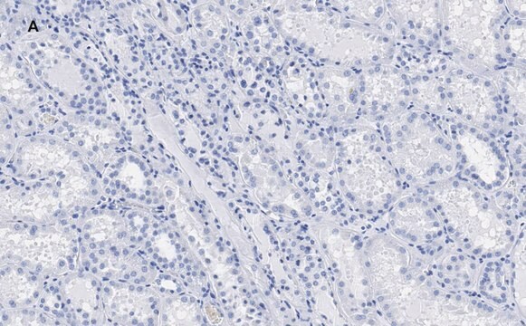
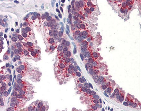
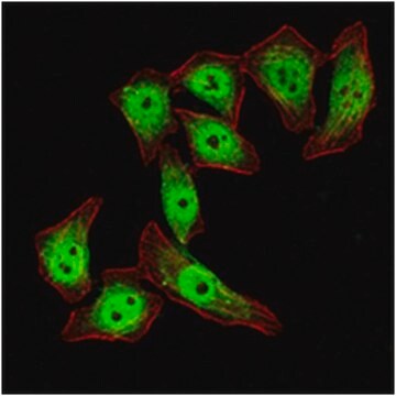
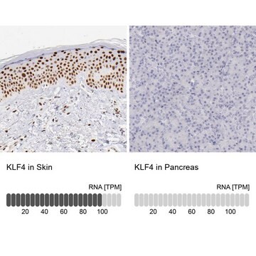
Anti-KLF4 Antibody is a Rabbit Polyclonal Antibody for detection of KLF4 also known as Kruppel-like Factor 4 & has been validated in ICC.
Immunocytochemistry: 1/200 to 1/1000.
Optimal dilutions must be determined by the user.
Optimal dilutions must be determined by the user.
Research Category
Epigenetics & Nuclear Function
Epigenetics & Nuclear Function
Research Sub Category
Transcription Factors
Transcription Factors
물리적 형태
Affinity purified immunoglobulin precipitated in a solution of 50% saturated ammonium sulfate and PBS containing no preservatives.
저장 및 안정성
Maintain unopened vial at -20°C for up to 6 months. Avoid repeated freeze/thaw cycles.
The rehydrated antibody solutions can be stored undiluted at 2-8°C for 2 months without any significant loss of activity. Note, the solution is not sterile, thus care should be taken if product is stored at 2-8°C.
For storage at -20°C, the addition of an equal volume of glycerol can be used, however, it is recommended that ACS grade or higher glycerol be used, as significant loss of activity can occur if the glycerol used is not of high quality.
For freezing, it is recommended that the rehydrated antibody solution be further diluted 1:1 with a 2% BSA (fraction V, highest-grade available) solution made with the rehydration buffer. The resulting 1% BSA/antibody solution can be aliquoted and stored frozen at -70°C for up to 6 months. Avoid repeated freeze/thaw cycles.
PREPARATION AND USE:
To reconstitute the antibody, centrifuge the antibody vial at moderate speed (5,000 rpm) for 5 minutes to pellet the precipitated antibody product. Carefully remove the ammonium sulfate/PBS buffer solution and discard. It is not necessary to remove all of the ammonium sulfate/PBS solution: 10 μL of residual ammonium sulfate solution will not effect the resuspension of the antibody. Do not let the protein pellet dry, as severe loss of antibody reactivity can occur.
Resuspend the antibody pellet in any suitable biological buffer, standard PBS or TBS (pH 7.3-7.5) are typical. Volumes required are not critical but it is suggested that the final antibody concentration be between 0.1 mg/mL and 1.0 mg/mL. For example, to achieve a1 mg/mL concentration with 50 μg of precipitated antibody, the amount of buffer needed would be 50 μL.
Carefully add the liquid buffer to the pellet. DO NOT VORTEX. Mix by gentle stirring with a wide pipet tip or gentle finger-tapping. Let the precipitated antibody rehydrate for 1 hour at 4-25°C prior to use. Small particles of precipitated antibody that fail to resuspend are normal. Vials are overfilled to compensate for any losses.
법적 정보
CHEMICON is a registered trademark of Merck KGaA, Darmstadt, Germany
면책조항
Unless otherwise stated in our catalog or other company documentation accompanying the product(s), our products are intended for research use only and are not to be used for any other purpose, which includes but is not limited to, unauthorized commercial uses, in vitro diagnostic uses, ex vivo or in vivo therapeutic uses or any type of consumption or application to humans or animals.
적합한 제품을 찾을 수 없으신가요?
당사의 제품 선택기 도구.을(를) 시도해 보세요.
시험 성적서(COA)
제품의 로트/배치 번호를 입력하여 시험 성적서(COA)을 검색하십시오. 로트 및 배치 번호는 제품 라벨에 있는 ‘로트’ 또는 ‘배치’라는 용어 뒤에서 찾을 수 있습니다.
Klf4 cooperates with Oct3/4 and Sox2 to activate the Lefty1 core promoter in embryonic stem cells.
Nakatake, Y; Fukui, N; Iwamatsu, Y; Masui, S; Takahashi, K; Yagi, R; Yagi, K; Miyazaki et al.
Molecular and cellular biology null
Sarah J Hainer et al.
Cell reports, 13(1), 61-69 (2015-09-29)
Functional interactions between gene regulatory factors and chromatin architecture have been difficult to directly assess. Here, we use micrococcal nuclease (MNase) footprinting to probe the functions of two chromatin-remodeling complexes. By simultaneously quantifying alterations in small MNase footprints over the
Benjamin Koopmansch et al.
Biochemical and biophysical research communications, 431(4), 652-657 (2013-02-05)
E-cadherin expression is repressed by ZEB2/SIP1 while it is induced by KLF4. Independent data from the literature indicate that these two transcription factors could bind close to each other in the proximal region of the E-cadherin gene promoter. We have
Long Wang et al.
Cytotechnology, 66(5), 729-740 (2013-10-05)
Human induced pluripotent stem (iPS) cells have great value for regenerative medicine, but are facing problems of low efficiency. MicroRNAs are a recently discovered class of 19-25 nt small RNAs that negatively target mRNAs. miR302/367 cluster has been demonstrated to
자사의 과학자팀은 생명 과학, 재료 과학, 화학 합성, 크로마토그래피, 분석 및 기타 많은 영역을 포함한 모든 과학 분야에 경험이 있습니다..
고객지원팀으로 연락바랍니다.