AB2226
Anti-MDGA1 Antibody
from rabbit, purified by affinity chromatography
동의어(들):
MAM domain-containing glycosylphosphatidylinositol anchor protein 1, GPI and MAM protein, GPIM, Glycosylphosphatidylinositol-MAM, MAM domain-containing protein 3
로그인조직 및 계약 가격 보기
모든 사진(1)
About This Item
UNSPSC 코드:
12352203
eCl@ss:
32160702
NACRES:
NA.41
추천 제품
생물학적 소스
rabbit
Quality Level
항체 형태
affinity isolated antibody
항체 생산 유형
primary antibodies
클론
polyclonal
정제법
affinity chromatography
종 반응성
mouse, rat
종 반응성(상동성에 의해 예측)
human (based on 100% sequence homology), bovine (based on 100% sequence homology)
기술
western blot: suitable
NCBI 수납 번호
UniProt 수납 번호
배송 상태
wet ice
타겟 번역 후 변형
unmodified
유전자 정보
human ... MDGA1(266727)
일반 설명
Studies report that MDGA1 is a layer-specific marker and an area-specific marker, being expressed in layers 2/3 throughout the neocortex, but within the primary somatosensory area (S1), MDGA1 is also uniquely expressed in layers 4 and 6a. Comparisons with other markers, including cadherins, serotonin, cytochrome oxidase, RORß, and COUP-TF1, reveal unique features of patterned expression of MDGA1 within cortex and S1 barrels. Furthermore, during early stages of development, MDGA1 is expressed by Reelin- and Tbr1-positive Cajal–Retzius neurons that originate from multiple sources outside of neocortex and emigrate into it. At even earlier stages, MDGA1 is expressed by the earliest diencephalic and mesencephalic neurons, which appear to migrate from a MDGA1-positive domain of progenitors in the diencephalon and form a "preplate." These findings show that MDGA1 is a unique marker for studies of cortical lamination and area patterning and together with recent reports suggest that MDGA1 has critical functions in forebrain/midbrain development.
특이성
This antibody recognizes the MAM domain of MDGA1.
면역원
Epitope: MAM domain
KLH-conjugated linear peptide corresponding to the MAM domain of MDGA1.
애플리케이션
Anti-MDGA1 Antibody detects level of MDGA1 & has been published & validated for use in Western Blotting.
Research Category
Neuroscience
Neuroscience
Research Sub Category
Developmental Neuroscience
Developmental Neuroscience
품질
Evaluated by Western Blot in untreated and PNGase F treated mouse P7 brain tissue lysate.
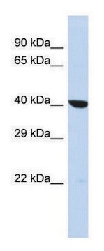
Western Blot Analysis: 0.5 µg/mL of this antibody detected MDGA1 in 5 µg of untreated and PNGase F treated mouse P7 brain tissue lysate. This antibody recognizes glycosolated (>160 kDa) (lane 1) and deglycosolated MDGA1 (lane 2).
Western Blot Analysis: 0.5 µg/mL of this antibody detected MDGA1 in 5 µg of untreated and PNGase F treated mouse P7 brain tissue lysate. This antibody recognizes glycosolated (>160 kDa) (lane 1) and deglycosolated MDGA1 (lane 2).
표적 설명
~130kDa observed. UniProt describes 2 isoforms produced by alternative splicing at ~106 kDa (Isoform 1), ~107 kDa (Isoform 2). An uncharacterized band may be observed at ~48 kDa in some tissue lysates.
물리적 형태
Affinity purified
Purified rabbit polyclonal in buffer containing 0.1 M Tris-Glycine (pH 7.4), 150 mM NaCl with 0.05% sodium azide.
저장 및 안정성
Stable for 1 year at 2-8°C from date of receipt.
분석 메모
Control
Untreated and PNGase F treated mouse P7 brain tissue lysate.
Untreated and PNGase F treated mouse P7 brain tissue lysate.
기타 정보
Concentration: Please refer to the Certificate of Analysis for the lot-specific concentration.
면책조항
Unless otherwise stated in our catalog or other company documentation accompanying the product(s), our products are intended for research use only and are not to be used for any other purpose, which includes but is not limited to, unauthorized commercial uses, in vitro diagnostic uses, ex vivo or in vivo therapeutic uses or any type of consumption or application to humans or animals.
적합한 제품을 찾을 수 없으신가요?
당사의 제품 선택기 도구.을(를) 시도해 보세요.
Storage Class Code
12 - Non Combustible Liquids
WGK
WGK 1
Flash Point (°F)
Not applicable
Flash Point (°C)
Not applicable
시험 성적서(COA)
제품의 로트/배치 번호를 입력하여 시험 성적서(COA)을 검색하십시오. 로트 및 배치 번호는 제품 라벨에 있는 ‘로트’ 또는 ‘배치’라는 용어 뒤에서 찾을 수 있습니다.
자사의 과학자팀은 생명 과학, 재료 과학, 화학 합성, 크로마토그래피, 분석 및 기타 많은 영역을 포함한 모든 과학 분야에 경험이 있습니다..
고객지원팀으로 연락바랍니다.