
추천 제품


생물학적 소스
rabbit
Quality Level
항체 형태
affinity purified immunoglobulin
항체 생산 유형
primary antibodies
클론
polyclonal
종 반응성
human, mouse, rat, bovine
제조업체/상표
Chemicon®
기술
ELISA: suitable
immunocytochemistry: suitable
immunohistochemistry (formalin-fixed, paraffin-embedded sections): suitable
immunoprecipitation (IP): suitable
western blot: suitable
NCBI 수납 번호
UniProt 수납 번호
배송 상태
dry ice
타겟 번역 후 변형
unmodified
유전자 정보
human ... SYN1(6853)
일반 설명
The synapsins are a family of proteins that have long been implicated in the regulation of neurotransmitter release at synapses. Specifically, they are thought to be involved in regulating the number of synaptic vesicles available for release via exocytosis at any one time. Synapsins are encoded by three different genes, synapsin I, II and III, and different neuron terminals will encode different amounts of these; synapsin will make up 1% of total brain protein at any one time.
Current studies suggest the following hypothesis for the role of synapsin: synapsins bind synaptic vesicles to components of the cytoskeleton which prevents them from migrating to the presynaptic membrane and releasing transmitter. During an action potential, synapsins are phosphorylated by Ca2+/calmodulin-dependent protein kinase II, releasing the synaptic vesicles and allowing them to move to the membrane and release their neurotransmitter.
Current studies suggest the following hypothesis for the role of synapsin: synapsins bind synaptic vesicles to components of the cytoskeleton which prevents them from migrating to the presynaptic membrane and releasing transmitter. During an action potential, synapsins are phosphorylated by Ca2+/calmodulin-dependent protein kinase II, releasing the synaptic vesicles and allowing them to move to the membrane and release their neurotransmitter.
특이성
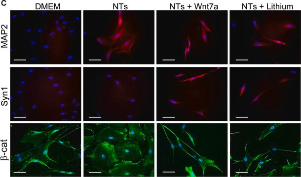
Synapsin I (Mr of 80,000 and 77,000). Synapsin is located only in brain nerve terminal and is an excellent marker for synapes. Immunolabeling is blocked by preadsorption of the antibody with synapsin I.
면역원
Synapsin I (mixture of Ia & Ib) purified from bovine brain.
애플리케이션
Research Category
Neuroscience
Neuroscience
Research Sub Category
Synapse & Synaptic Biology
Synapse & Synaptic Biology
This Anti-Synapsin I Antibody is validated for use in ELISA, IC, IH, IH(P), IP, WB for the detection of Synapsin I.
Western blotting:
1:200-1:1,000 dilution of a previous lot was used.
Immunocytochemistry:
1:500-1:2,000 dilution of a previous lot was used.
Immunoprecipitation:
1 μg of a previous lot immunoprecipitated all of the synapsin I from a SDS homogenate of 200 μg of rat brain protein.
Note: The above dilutions are with 35S-protein A; with ECL dilutions may need to be considerably higher to obtain specific immunolabeling.
Immunohistochemistry:
1:500-1:2,500
ELISA:
1:2,500-1:10,00
Optimal working dilutions must be determined by the end user.
품질
Immunohistochemistry(paraffin):
Synapsin I (AB1543P) representative staining pattern/morphology in rat hippocampal neurons. Tissue was pretreated with Citrate pH 6.0, antigen retrieval. This lot of antibody was diluted to 1:1000, IHC-Select reagents used with HRP-DAB. Immunoreactivity is seen directly associated with terminal end of neuronal cell body.
IHC-Paraffin Staining With Epitope Retrieval:
Rat Hippocampus
표적 설명
77 & 80 kDa
결합
Replaces: MAB355
물리적 형태
Format: Purified
ImmunoAffinity Purified
Purified rabbit polyclonal in buffer lyophilized from 5 mM ammonium bicarbonate. Reconstitute with 50 μL of PBS.
저장 및 안정성
Stable for 1 year at -20ºC from date of receipt.
분석 메모
Control
Brain tissue.
Brain tissue.
기타 정보
Concentration: Please refer to the Certificate of Analysis for the lot-specific concentration.
법적 정보
CHEMICON is a registered trademark of Merck KGaA, Darmstadt, Germany
면책조항
Unless otherwise stated in our catalog or other company documentation accompanying the product(s), our products are intended for research use only and are not to be used for any other purpose, which includes but is not limited to, unauthorized commercial uses, in vitro diagnostic uses, ex vivo or in vivo therapeutic uses or any type of consumption or application to humans or animals.
적합한 제품을 찾을 수 없으신가요?
당사의 제품 선택기 도구.을(를) 시도해 보세요.
Storage Class Code
11 - Combustible Solids
WGK
WGK 1
시험 성적서(COA)
제품의 로트/배치 번호를 입력하여 시험 성적서(COA)을 검색하십시오. 로트 및 배치 번호는 제품 라벨에 있는 ‘로트’ 또는 ‘배치’라는 용어 뒤에서 찾을 수 있습니다.
M P Cid et al.
Neuroscience, 189, 337-344 (2011-06-04)
We previously found that the glutamate release was decreased in synaptosomes from rat cerebral cortex during the development of experimental autoimmune encephalomyelitis (EAE), the animal model of multiple sclerosis. Various other reports have shown a deficit in the expression of
LRRTMs and neuroligins bind neurexins with a differential code to cooperate in glutamate synapse development.
Siddiqui TJ, Pancaroglu R, Kang Y, Rooyakkers A, Craig AM
The Journal of Neuroscience null
Ling Li et al.
PloS one, 5(7), e11503-e11503 (2010-07-17)
Proper function of the mammalian brain relies on the establishment of highly specific synaptic connections among billions of neurons. To understand how complex neural circuits function, it is crucial to precisely describe neuronal connectivity and the distributions of synapses to
Diffuse and specific tectopulvinar terminals in the tree shrew: synapses, synapsins, and synaptic potentials.
Wei, H; Masterson, SP; Petry, HM; Bickford, ME
Testing null
Eric Wersinger et al.
PloS one, 5(11), e13836-e13836 (2010-11-17)
Outer hair cells are the specialized sensory cells that empower the mammalian hearing organ, the cochlea, with its remarkable sensitivity and frequency selectivity. Sound-evoked receptor potentials in outer hair cells are shaped by both voltage-gated K(+) channels that control the
자사의 과학자팀은 생명 과학, 재료 과학, 화학 합성, 크로마토그래피, 분석 및 기타 많은 영역을 포함한 모든 과학 분야에 경험이 있습니다..
고객지원팀으로 연락바랍니다.