
추천 제품


생물학적 소스
rabbit
Quality Level
항체 형태
affinity isolated antibody
항체 생산 유형
primary antibodies
클론
polyclonal
정제법
affinity chromatography
종 반응성
mouse
종 반응성(상동성에 의해 예측)
canine (based on 100% sequence homology), canine, bovine, opossum, horse, rat, pig
기술
immunohistochemistry: suitable (paraffin)
western blot: suitable
NCBI 수납 번호
UniProt 수납 번호
배송 상태
wet ice
타겟 번역 후 변형
unmodified
유전자 정보
human ... TBR1(10716)
일반 설명
T-box brain protein 1 (UniProt: Q64336; also known as T-brain-1, TBR-1, TES-56) is encoded by the Tbr1 gene (Gene ID: 21375) in murine species. Tbr1 is a homodimeric nuclear protein that serves as a transcriptional repressor and is involved in multiple aspects of cortical development, including neuronal migration, laminar and areal identity, and axonal projection. As transcriptional repressor of FEZ Family Zinc Finger 2 (FEZF2), it blocks the formation of the corticospinal (CS) tract from layer 6 projection neurons, thereby restricting the origin of CS axons specifically to layer 5 neurons. Its T-box DNA binding domain is localized in amino acids 213-393. Its expression is observed in developing and adult cerebral cortex and in olfactory bulbs. It is detected around day 10 of embryonic development in the preplate and at day 12.5 in the cortical plate. It is highly expressed in glutamatergic early-born cortical neurons. Tbr1 null mice exhibit severe defects of frontal cortex and layer 6 differentiation. (Ref.: Han, W., et al. (2011). Proc. Natl. Acad. Sci. USA. 108(7); 3041-3046; Bedogni, F., et al (2010). Proc. Natl Acad. Sci. USA. 107(29); 13129-13134; Hevner, RF., et al. (2001). Neuron. 29(2); 353-366).
특이성
This rabbit polyclonal antibody detects T-box brain protein 1 (Tbr1).
면역원
KLH-conjugated linear peptide corresponding to 18 amino acids from the N-terminal region of mouse T-box brain protein 1 (Tbr1).
KLH-conjugated linear peptide corresponding to mouse Tbr1 near the N-terminus.
애플리케이션
Quality Control Testing
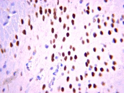
Evaluated by Immunohistochemistry (Paraffin) in Mouse brain tissue sections.Immunohistochemistry (Paraffin) Analysis: A 1:400 dilution of this antibody detected Tbr1 in Mouse cerebral cortex and cerebellum tissue sections.
Tested Applications
Western Blotting Analysis: A 1:500 dilution from a representative lot detected Tbr1 in Mouse fetal brain tissue lysate.Note: Actual optimal working dilutions must be determined by end user as specimens, and experimental conditions may vary with the end user.
품질
Evaluated by Immunohistochemistry in mouse frontal cortex tissue.
Immunohistochemistry Analysis: 1:400 dilution of this antibody detected Tbr1 in mouse frontal cortex tissue.
Immunohistochemistry Analysis: 1:400 dilution of this antibody detected Tbr1 in mouse frontal cortex tissue.
표적 설명
~76 kDa observed; 73.94 kDa calculated. Uncharacterized bands may be observed in some lysate(s).
물리적 형태
Affinity purified
Purified rabbit polyclonal antibody in buffer containing 0.1 M Tris-Glycine (pH 7.4), 150 mM NaCl with 0.05% sodium azide.
저장 및 안정성
Recommended storage: +2°C to +8°C.
분석 메모
Control
Mouse frontal cortex tissue
Mouse frontal cortex tissue
기타 정보
Concentration: Please refer to the Certificate of Analysis for the lot-specific concentration.
면책조항
Unless otherwise stated in our catalog or other company documentation accompanying the product(s), our products are intended for research use only and are not to be used for any other purpose, which includes but is not limited to, unauthorized commercial uses, in vitro diagnostic uses, ex vivo or in vivo therapeutic uses or any type of consumption or application to humans or animals.
적합한 제품을 찾을 수 없으신가요?
당사의 제품 선택기 도구.을(를) 시도해 보세요.
Storage Class Code
12 - Non Combustible Liquids
WGK
WGK 1
Flash Point (°F)
Not applicable
Flash Point (°C)
Not applicable
시험 성적서(COA)
제품의 로트/배치 번호를 입력하여 시험 성적서(COA)을 검색하십시오. 로트 및 배치 번호는 제품 라벨에 있는 ‘로트’ 또는 ‘배치’라는 용어 뒤에서 찾을 수 있습니다.
Epigenomic Analysis of Multilineage Differentiation of Human Embryonic Stem Cells.
Xie, Wei, et al.
Cell (2013)
J Ni et al.
International journal of molecular medicine, 35(6), 1755-1760 (2015-04-08)
Knee osteoarthritis (OA) is the most prevalent type of OA and the cytokine, oncostatin M (OSM), may contribute to the pathogenesis of OA. However, the exact role of OSM in the development of knee OA and the underlying mechanisms are
Xue Li et al.
Cerebral cortex (New York, N.Y. : 1991), 30(7), 3960-3976 (2020-02-03)
De novo microdeletion of chromosome 2p15-16.1 presents clinically recognizable phenotypes that include mental retardation, autism, and microcephaly. Chromosomal maintenance 1 (CRM1) is a gene commonly missing in patients with 2p15-16.1 microdeletion and one of two genes found in the smallest
Christine Sauerland et al.
Cerebral cortex (New York, N.Y. : 1991), 28(1), 145-157 (2017-12-19)
A hallmark of mammalian brain evolution is the emergence of the neocortex, which has expanded in all mammalian infraclasses (Eutheria, Marsupialia, Monotremata). In eutherians, neocortical neurons derive from distinct neural stem and progenitor cells (NPCs). However, precise data on the
Ilaria Favicchia et al.
Frontiers in molecular neuroscience, 14, 663598-663598 (2021-09-24)
Tbx1 mutant mice are a widely used model of 22q11.2 deletion syndrome (22q11.2DS) because they manifest a broad spectrum of physical and behavioral abnormalities that is similar to that found in 22q11.2DS patients. In Tbx1 mutants, brain abnormalities include changes
자사의 과학자팀은 생명 과학, 재료 과학, 화학 합성, 크로마토그래피, 분석 및 기타 많은 영역을 포함한 모든 과학 분야에 경험이 있습니다..
고객지원팀으로 연락바랍니다.