
추천 제품


양식
liquid
Quality Level
사용
sufficient for ≤2,500 tests
포장
pkg of 1 kit
제조업체/상표
Roche
저장 조건
protect from light
기술
tissue culture: suitable
λmax
450-500 nm
응용 분야
cell analysis
detection
검출 방법
colorimetric
저장 온도
−20°C
일반 설명
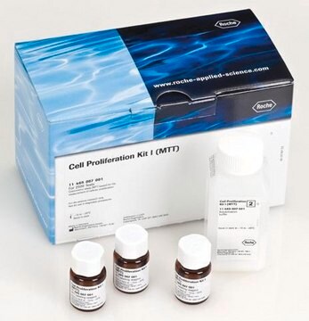
The Cell Proliferation Kit II (XTT) is a colorimetric assay for the nonradioactive quantification of cellular proliferation, viability, and cytotoxicity. Sample material is either adherent or suspension cells cultured in 96-well microplates.
Colorimetric assays analyze the number of viable cells by the cleavage of tetrazolium salts added to the culture medium. This technique requires neither washing nor harvesting of cells, and the complete assay, from microculture to data analysis by an ELISA reader, is performed in the same microplate.
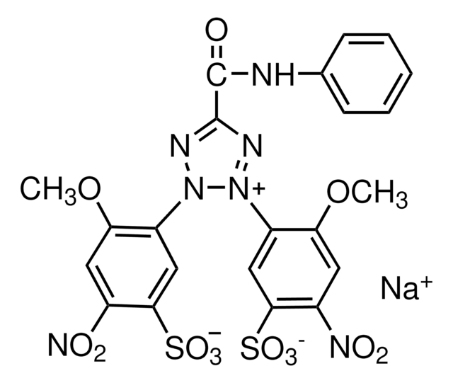
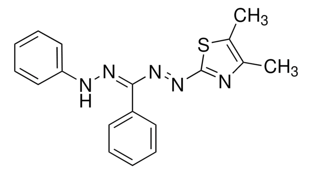
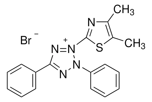
More recently, the tetrazolium salt XTT was described. In contrast to MTT, the cleavage product of XTT is soluble in water; therefore, a solubilization step is not required. The tetrazolium salt XTT is cleaved to formazan by a complex cellular mechanism. This bioreduction occurs in viable cells only, and is related to NAD(P)H production through glycolysis. Therefore, the amount of formazan dye formed directly correlates to the number of metabolically active cells in the culture.
Colorimetric assays analyze the number of viable cells by the cleavage of tetrazolium salts added to the culture medium. This technique requires neither washing nor harvesting of cells, and the complete assay, from microculture to data analysis by an ELISA reader, is performed in the same microplate.
More recently, the tetrazolium salt XTT was described. In contrast to MTT, the cleavage product of XTT is soluble in water; therefore, a solubilization step is not required. The tetrazolium salt XTT is cleaved to formazan by a complex cellular mechanism. This bioreduction occurs in viable cells only, and is related to NAD(P)H production through glycolysis. Therefore, the amount of formazan dye formed directly correlates to the number of metabolically active cells in the culture.
애플리케이션
The Cell Proliferation Kit II (XTT) is used for the nonradioactive, spectrophotometric quantification of cell proliferation and viability in cell populations using the 96-well-plate format. It can be used for:
- Measurement of cell proliferation in response to growth factors, cytokines, mitogens, and nutrients.
- Analysis of cytotoxic and cytostatic compounds, such as anti-cancer drugs and other pharmaceutical compounds.
- Assessment of growth-inhibitory antibodies and physiological mediators that inhibit cell growth.
- Testing of biocompatibility of various scaffolds, employed in bone tissue engineering, for bone cell growth.
- cell viability assay.
특징 및 장점
- Safe and easy: Eliminate radioactive isotopes, washing steps, and additional reagents.
- Accurate: The absorbance obtained strongly correlates to the cell number.
- Sensitive: Detect low cell numbers.
- Fast: Process a large number of samples using a multi-well ELISA reader.
포장
1 kit containing 2 components.
원리
The assay is based on the cleavage of the tetrazolium salt XTT in the presence of an electron-coupling reagent, producing a soluble formazan salt. This conversion only occurs in viable cells. Cells grown in a 96-well tissue culture plate are incubated with the XTT labeling mixture for 2 - 20 hours. After this incubation period, the formazan dye formed is quantitated using a scanning multi-well spectrophotometer (ELISA reader). The measured absorbance directly correlates to the number of viable cells.
Cell proliferation and viability assays are of particular importance for routine applications in cell biology. Tetrazolium salts (e.g., MTT, XTT, WST-1) are particularly useful for this type of analysis. Tetrazolium salts are cleaved to formazan by the succinate-tetrazolium reductase system (EC 1.3.99.1) which belongs to the respiratory chain of the mitochondria, and is only active in metabolically intact cells.
Cell proliferation and viability assays are of particular importance for routine applications in cell biology. Tetrazolium salts (e.g., MTT, XTT, WST-1) are particularly useful for this type of analysis. Tetrazolium salts are cleaved to formazan by the succinate-tetrazolium reductase system (EC 1.3.99.1) which belongs to the respiratory chain of the mitochondria, and is only active in metabolically intact cells.
제조 메모
Working solution: Preparation of solutions
Thaw XTT labeling reagent and electron-coupling reagent, respectively in a water bath at 37 °C. Mix each vial thoroughly to obtain a clear solution.
XTT labeling mixture
To perform a cell proliferation assay (XTT) with one microplate (96 wells) mix 5 ml XTT labeling reagent with 0.1 ml electron coupling reagent.
Note: To obtain reliable results thaw and mix XTT labeling reagent and electron coupling reagent immediately before use.
Working instruction
Cells are grown in microplates (tissue culture grade, 96 wells, flat bottom) in a final volume of 100 μl culture medium per well, according to the media needs of the cells in a humidified atmosphere (e.g., 37 °C, 6.5% CO2).
The incubation period of the cell cultures depends on the particular experimental approach and on the cell line, used for the assay. For most experimental setups, the incubation of cells for 24 to 96 hours is appropriate.
After the incubation period, add to each well 50 μl of the XTT labeling mixture, prepared as described above (final XTT concentration 0.3 mg/ml).
Incubate the microplate for 4 to 24 hours in a humidified atmosphere (e.g., 37 °C, 6.5% CO2).
Note: The incubation time varies with the individual experimental setup (e.g., cell type and cell concentration, used). Therefore, we recommend to measure the absorption as described at different time points after addition of XTT labeling mixture (e.g., 4, 6, 8, 12, and 18 hours) using one and the same microplate to determine the optimal incubation period for the particular experimental setup.
Storage conditions (working solution): Thaw reagents immediately before use. It is recommended to prepare appropriate aliquots [5 ml XTT labeling reagent and 0.1 ml electron coupling reagent are required for the performance of the assay with one microplate (96 wells)]
Note: Avoid repeated thawing and freezing.
기타 정보
For life science research only. Not for use in diagnostic procedures.
키트 구성품 전용
제품 번호
설명
- XTT Labeling Reagent
- Electron-coupling Reagent
Storage Class Code
12 - Non Combustible Liquids
WGK
nwg
Flash Point (°F)
does not flash
Flash Point (°C)
does not flash
이미 열람한 고객

A combination of photodynamic therapy and chemotherapy displays a differential cytotoxic effect on human metastatic melanoma cells.
Nsole Biteghe F A and Davids L M
Journal of Photochemistry and Photobiology. B, Biology, 166, 18-27 (2017)
High-Temperature Requirement A1 (Htra1) - A Novel Regulator of Canonical Wnt Signaling.
Oriane G, et al.
Scientific Reports, 7(1), 17995-17995 (2017)
Senescent growth arrest in mesenchymal stem cells is bypassed by Wip1-mediated downregulation of intrinsic stress signaling pathways.
Lee J S, et al.
Stem Cells, 27(8), 1963-1975 (2009)
Eun Young Park et al.
The Journal of biological chemistry, 289(13), 9254-9262 (2014-02-12)
Autosomal dominant polycystic kidney disease (ADPKD) is the most common inherited renal disorder. Although a myriad of research groups have attempted to identify a new therapeutic target for ADPKD, no drug has worked well in clinical trials. Our research group
Constantin Heil
Open biology, 9(1), 180145-180145 (2019-04-09)
Cerebellar granule cell progenitors (GCPs) undergo proliferation in the post-natal cerebellum that is dependent on sonic hedgehog (SHH) signalling. Deregulated SHH signalling leads to type 2 medulloblastoma (MB). In this work, a novel cell culture protocol is described, which is
문서
Cell based assays for cell proliferation (BrdU, MTT, WST1), cell viability and cytotoxicity experiments for applications in cancer, neuroscience and stem cell research.
프로토콜
Protocol Guide: XTT Assay for Cell Viability and Proliferation
자사의 과학자팀은 생명 과학, 재료 과학, 화학 합성, 크로마토그래피, 분석 및 기타 많은 영역을 포함한 모든 과학 분야에 경험이 있습니다..
고객지원팀으로 연락바랍니다.