Skip to Content
Merck
All Photos(2)

Key Documents

Safety Information

A4335

Sigma-Aldrich

Monoclonal Anti-Myosin (Skeletal, Fast)−Alkaline Phosphatase antibody produced in mouse

clone MY-32, purified from hybridoma cell culture

Synonym(s):

Monoclonal Anti-Myosin (Skeletal, Fast) antibody produced in mouse

Sign Into View Organizational & Contract Pricing


About This Item

MDL number:
UNSPSC Code:
12352203
NACRES:
NA.41

biological source

mouse

Quality Level

conjugate

alkaline phosphatase conjugate

antibody form

purified immunoglobulin

antibody product type

primary antibodies

clone

MY-32, monoclonal

form

buffered aqueous glycerol solution

species reactivity

rat, chicken, rabbit, mouse, human, bovine, guinea pig, feline

technique(s)

direct immunofluorescence: 1:150 using formalin-fixed, paraffin-embedded human or animal skeletal muscle sections

isotype

IgG1

UniProt accession no.

shipped in

wet ice

storage temp.

2-8°C

target post-translational modification

unmodified

Gene Information

Looking for similar products? Visit Product Comparison Guide

General description

Localizes an epitope on the myosin heavy chain. Stains the fast (type II) and neonatal isomyosin molecules found in skeletal muscle, but does not stain cardiac muscle, smooth muscle or non-muscle myosin in cultured cells. Does react with human rhabdomyosarcomas.
Monoclonal Anti-Myosin (Skeletal, Fast) (mouse IgG1 isotype) is derived from the hybridoma produced by the fusion of mouse myeloma cells and splenocytes from an immunized mouse.

Specificity

Monoclonal Anti-Myosin (Skeletal, Fast) specifically binds to an epitope in adult rat skeletal myosin heavy chains IIa, IIb and IId (IIx) (in fast-twitch fibers)†. The antibody product is also reactive in humans, bovines, cats, rabbits, mice, guinea pigs and chickens.

Immunogen

rabbit muscle myosin.

Application

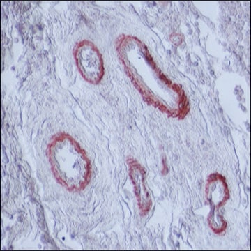
Monoclonal Anti-Myosin (Skeletal, Fast) Alkaline Phosphatase antibody produced in mouse has been used for the detection and localization of skeletal muscle fast and neonatal myosins using immunohistochemistry. It may be used in muscle fiber typing, in studies of in vivo and in vitro muscle development and in the diagnosis of rhabdomyosarcomas.
The level of mysosin (fast) in serum samples from sportsmen with past injury was determined by western blot using monoclonal mouse anti-myosin (skeletal/fast) as the primary antibody at a dilution of 1:90000.

Biochem/physiol Actions

Myosin (Skeletal, Fast) is useful not only in fiber typing but also in detection of myogenic tumors. It enables retrospective as well as prospective fiber typing using formalin-fixed, paraffin-embedded material.
Myosins are motor proteins that interact with actin filaments to regulate cell movement. Myosins also modulate cell shape and signaling. Skeletal muscle myosins have three ′fast′ myosin heavy chains isoforms (IIa, IIx, and IIb), a ′slow′ beta-myosin heavy chain isoform and three major myosin light-chain (MLC) isoforms. These myosin isoforms regulate skeletal muscle shortening.

Physical form

Solution in 0.05 M Tris buffer, pH 8.0, containing 1% bovine serum albumin, 1 mM MgCl2, 50% glycerol, and 15 mM sodium azide.

Disclaimer

Unless otherwise stated in our catalog or other company documentation accompanying the product(s), our products are intended for research use only and are not to be used for any other purpose, which includes but is not limited to, unauthorized commercial uses, in vitro diagnostic uses, ex vivo or in vivo therapeutic uses or any type of consumption or application to humans or animals.

Not finding the right product?  

Try our Product Selector Tool.

Storage Class Code

10 - Combustible liquids

WGK

WGK 2


Regulatory Listings

Regulatory Listings are mainly provided for chemical products. Only limited information can be provided here for non-chemical products. No entry means none of the components are listed. It is the user’s obligation to ensure the safe and legal use of the product.

JAN Code

A4335-VAR:
A4335-BULK:
A4335-.2ML:


Certificates of Analysis (COA)

Search for Certificates of Analysis (COA) by entering the products Lot/Batch Number. Lot and Batch Numbers can be found on a product’s label following the words ‘Lot’ or ‘Batch’.

Already Own This Product?

Find documentation for the products that you have recently purchased in the Document Library.

Visit the Document Library

F Russell Westwood et al.
Toxicologic pathology, 36(2), 345-352 (2008-03-26)
Rosuvastatin is a relatively new member of the statin family (HMG-CoA reductase inhibitors), with superior lipid-lowering effects and a pattern of clinical side effects, including a low incidence of myopathy, similar to other widely prescribed statins. This article describes investigations
Christopher Taber et al.
Sports (Basel, Switzerland), 6(2) (2018-06-19)
The purpose of this study was to examine the effects of a recovery supplement compared with a placebo on muscle morphology in trained weightlifters. Vastus lateralis and muscle fiber cross sectional area of type I and type II fibers were
Katy C Liu et al.
Bioarchitecture, 2(5), 158-170 (2012-09-08)
The development of cell-cell junctions was a fundamental step in metazoan evolution, and human health depends on the formation and function of cell junctions. Although it has long been known that actin and conventional myosin have important roles in cell
A J Harris et al.
Development (Cambridge, England), 107(4), 751-769 (1989-12-01)
The expression of myosin isoforms was studied during development of calf muscles in foetal and neonatal rats, using monoclonal antibodies against slow, embryonic and neonatal isoforms of myosin heavy chain (MHC). Primary myotubes had appeared in all prospective rat calf
Vivi F H Jensen et al.
Basic & clinical pharmacology & toxicology, 122(1), 165-175 (2017-08-18)
Peripheral neuropathy is one of the most common complications of diabetic hyperglycaemia. Insulin-induced hypoglycaemia (IIH) might potentially exacerbate or contribute to neuropathy as hypoglycaemia also causes peripheral neuropathy. In rats, IIH induces neuropathy associated with skeletal muscle changes. Aims of

Our team of scientists has experience in all areas of research including Life Science, Material Science, Chemical Synthesis, Chromatography, Analytical and many others.

Contact Technical Service