Skip to Content
Merck
All Photos(1)

Key Documents

Safety Information

M7786

Sigma-Aldrich

Monoclonal Anti-Myosin (Smooth) antibody produced in mouse

clone hSM-V, ascites fluid

Sign Into View Organizational & Contract Pricing


About This Item

MDL number:
UNSPSC Code:
12352203
NACRES:
NA.41

biological source

mouse

Quality Level

conjugate

unconjugated

antibody form

ascites fluid

antibody product type

primary antibodies

clone

hSM-V, monoclonal

mol wt

antigen 200-204 kDa

contains

15 mM sodium azide

species reactivity

guinea pig, human, pig, canine, rabbit, chicken

technique(s)

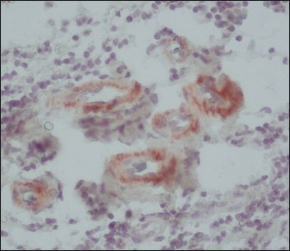
immunohistochemistry: 1:500 using and animal frozen section using acetone fixed human.
immunohistochemistry: suitable using using methacarn-fixed, paraffin-embedded sections of human and animal tissue
immunoprecipitation (IP): suitable
western blot: suitable

isotype

IgG1

UniProt accession no.

shipped in

dry ice

storage temp.

−20°C

target post-translational modification

unmodified

Gene Information

human ... MYH11(4629)

General description

Monoclonal Anti-Myosin (Smooth) (mouse IgG1 isotype) is derived from the hSM-V hybridoma produced by the fusion of mouse myeloma cells and splenocytes from BALB/c mice immunized with human uterus smooth muscle extract. Myosin (~500 kDa) is a cytoskeletal protein containing two identical heavy chains (~200 kDa each) and four light chains (15-26 kDa). Myosin molecules consist of two major regions: tail (rod) and heads.

Specificity

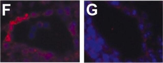
Mouse monoclonal clone hSM-V anti-Myosin (Smooth) antibody reacts in immunoblotting assays with myosin heavy chain polypeptides of 204 and 200 kDa (SM-1 and SM-2). It does not cross-react with skeletal, cardiac or non-muscle myosin. The antibody stains vascular and visceral smooth muscle cells, as well as cells that have smooth muscle-like characteristics (myofibroblasts and myoepithelial cells). It does not recognize epithelial, endothelial or connective tissue fibroblast cells. The product reacts with smooth muscle tissue from human, dog, rabbit, pig, guinea pig, and chicken.

Immunogen

human uterus smooth muscle extract.

Application

The antibody may be used in:
  • immunoblotting
  • immunoprecipitation
  • immunohistochemistry
  • immunocytochemistry
  • flow cytometry
  • immunofluorescence

Biochem/physiol Actions

Myosin implicated in cell motility. They aggregate into filaments through the tail region and interact with actin and with ATP through the head region. Myosin molecules spontaneously assemble into filaments in solutions of physiologic ionic strength and pH. In fact, the thick filament consists mainly of myosin molecules. The ATPase of myosin is activated by actin. This activation is the immediate source of the free energy that drives muscle contraction. It binds to the polymerized form of actin, the major constituent of the thin filament. Myosin is generally assumed to be a very specific and reliable smooth muscle marker, provided that the antibody used is specific for smooth muscle myosin and does not cross-react with skeletal, cardiac or non-muscle myosins.

Disclaimer

Unless otherwise stated in our catalog or other company documentation accompanying the product(s), our products are intended for research use only and are not to be used for any other purpose, which includes but is not limited to, unauthorized commercial uses, in vitro diagnostic uses, ex vivo or in vivo therapeutic uses or any type of consumption or application to humans or animals.

Not finding the right product?  

Try our Product Selector Tool.

Storage Class Code

10 - Combustible liquids

WGK

WGK 2

Flash Point(F)

Not applicable

Flash Point(C)

Not applicable


Regulatory Listings

Regulatory Listings are mainly provided for chemical products. Only limited information can be provided here for non-chemical products. No entry means none of the components are listed. It is the user’s obligation to ensure the safe and legal use of the product.

JAN Code

M7786-.2ML:
M7786-VAR:
M7786-.5ML:
M7786-100UL:
M7786-BULK:


Certificates of Analysis (COA)

Search for Certificates of Analysis (COA) by entering the products Lot/Batch Number. Lot and Batch Numbers can be found on a product’s label following the words ‘Lot’ or ‘Batch’.

Already Own This Product?

Find documentation for the products that you have recently purchased in the Document Library.

Visit the Document Library

Phosphorylation of the regulatory light chain of myosin in striated muscle: methodological perspectives
Yu H, et al.
European Biophysics Journal, 45(8), 779-805 (2016)
Endothelial-to-mesenchymal transition drives atherosclerosis progression
Chen PY, et al.
The Journal of Clinical Investigation, 125(12), 4514-4528 (2015)
Human vascular model with defined stimulation medium - a characterization study
Huttala O, et al.
ALTEX, Citations(may), include-include (2015)
Myosin: The Actin Motor Protein
Molecular Cell Biology. 4th edition. (2000)
Direct measurements of local coupling between myosin molecules are consistent with a model of muscle activation
Walcott S and Kad NM
PLoS Computational Biology, 11(11), e1004599-e1004599 (2015)

Our team of scientists has experience in all areas of research including Life Science, Material Science, Chemical Synthesis, Chromatography, Analytical and many others.

Contact Technical Service