Skip to Content
Merck
All Photos(2)

Documents

Safety Information

MABN794

Sigma-Aldrich

Anti-GLAST Antibody, clone 8C11.1

clone 8C11.1, from mouse

Synonym(s):

Excitatory amino acid transporter 1, Sodium-dependent glutamate/aspartate transporter 1, GLAST-1, Solute carrier family 1 member 3, GLAST

Sign Into View Organizational & Contract Pricing


About This Item

UNSPSC Code:
12352203
eCl@ss:
32160702
NACRES:
NA.43

biological source

mouse

Quality Level

antibody form

purified antibody

antibody product type

primary antibodies

clone

8C11.1, monoclonal

species reactivity

human, rat

technique(s)

immunohistochemistry: suitable (paraffin)
western blot: suitable

isotype

IgG1λ

NCBI accession no.

UniProt accession no.

shipped in

wet ice

target post-translational modification

unmodified

Gene Information

human ... SLC1A3(6507)

General description

Excitatory amino acid transporter 1 (UniProt P43003; also known as GLAST-1, Sodium-dependent glutamate/aspartate transporter 1, Solute carrier family 1 member 3) is encoded by the SLC1A3 (also known as EA6, EAAT1, GLAST, GLAST1) gene in human (Entrez Gene ID 6507). GLAST-1 is a multitransmembrane protein that functions as a high affinity glutamate transporter and mediates the termination of postsynaptic action of glutamate in central nervous system by rapidly removing released glutamate from the synaptic cleft. GLAST-1 also transports L- and D-aspartate. GLAST gene mutations are linked to episodic ataxia type 6 (EA6), a disorder characterized by seizures, migraine, and alternating hemiplegia. There are 5 known spliced variants. Isoform 2 has no functional glutamate uptake activity and exerts a dominant negative effect against isoform 1.

Specificity

Clone 8C11.1 epitope lies within the second extracellular segement of GLAST isoforms 1 & 2. The epitope region is also present in isoform 4, but not in isforms 3 & 5.

Immunogen

Epitope: Extracellular domain
GST-tagged recombinant protein corresponding to the extracellular domain of human GLAST.

Application

Anti-GLAST Antibody, clone 8C11.1 is an antibody against GLAST for use in Western Blotting, Immunohistochemistry .
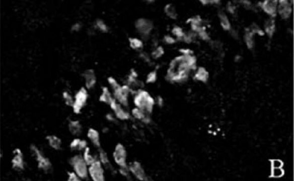
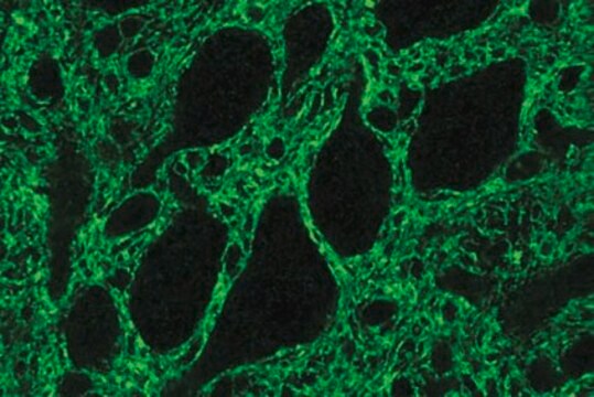
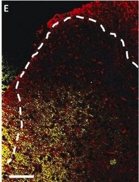
Immunohistochemistry Analysis: A 1:1,000 dilution from a representative lot detected GLAST in human cerebellum and rat cerebellum tissue.
Research Category
Neuroscience
Research Sub Category
Developmental Signaling

Quality

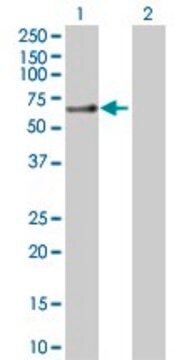
Evaluated by Western Blotting in human brain tissue lysate.

Western Blotting Analysis: 1.0 µg/mL of this antibody detected GLAST in 200 µg of human brain tissue lysate.

Target description

~55 kDa observed

Physical form

Format: Purified
Protein G Purified
Purified mouse monoclonal IgG1λ antibody in buffer containing 0.1 M Tris-Glycine (pH 7.4), 150 mM NaCl with 0.05% sodium azide.

Storage and Stability

Stable for 1 year at 2-8°C from date of receipt.

Other Notes

Concentration: Please refer to lot specific datasheet.

Disclaimer

Unless otherwise stated in our catalog or other company documentation accompanying the product(s), our products are intended for research use only and are not to be used for any other purpose, which includes but is not limited to, unauthorized commercial uses, in vitro diagnostic uses, ex vivo or in vivo therapeutic uses or any type of consumption or application to humans or animals.

Not finding the right product?  

Try our Product Selector Tool.

Storage Class Code

12 - Non Combustible Liquids

WGK

WGK 1

Flash Point(F)

Not applicable

Flash Point(C)

Not applicable


Regulatory Listings

Regulatory Listings are mainly provided for chemical products. Only limited information can be provided here for non-chemical products. No entry means none of the components are listed. It is the user’s obligation to ensure the safe and legal use of the product.

JAN Code

MABN794:


Certificates of Analysis (COA)

Search for Certificates of Analysis (COA) by entering the products Lot/Batch Number. Lot and Batch Numbers can be found on a product’s label following the words ‘Lot’ or ‘Batch’.

Already Own This Product?

Find documentation for the products that you have recently purchased in the Document Library.

Visit the Document Library

Haijun Zhang et al.
The journal of pain, 13(3), 293-303 (2012-01-31)
Paclitaxel often induces persistent painful neuropathy as its most common treatment-limiting side effect. Little is known concerning the underlying mechanisms. Given the prominent role of glial cells in many types of neuropathic pain, we investigated here the morphological and functional
Sandra Gómez-López et al.
Scientific reports, 6, 24028-24028 (2016-04-12)
Parkinson's disease (PD) is a multifactorial neurodegenerative disorder, characterised by the progressive loss of midbrain dopaminergic neurons and a variety of motor symptoms. The gene coding for the phospholipid phosphatase 3, PLPP3 (formerly PPAP2B or LPP3), maps within the PARK10
Rebeca Rosas-Hernández et al.
Journal of neurochemistry, 149(2), 242-254 (2018-12-28)
Alpha-amino-3-hydroxy-5-methylisoxazole-4-propionic acid glutamate receptors have been shown to modulate the morphology of the lamelar processes of Bergmann glia cells in the molecular layer of the cerebellum. Here we suggest that reorganization of F-actin may underlay the changes in the morphology

Our team of scientists has experience in all areas of research including Life Science, Material Science, Chemical Synthesis, Chromatography, Analytical and many others.

Contact Technical Service