Skip to Content
Merck
All Photos(1)

Key Documents

Safety Information

CBL271

Sigma-Aldrich

Anti-Fibroblasts Antibody, clone TE-7

clone TE-7, Chemicon®, from mouse

Synonym(s):

Fibroblast Marker

Sign Into View Organizational & Contract Pricing


About This Item

UNSPSC Code:
12352203
eCl@ss:
32160702
NACRES:
NA.41

biological source

mouse

Quality Level

antibody form

purified antibody

antibody product type

primary antibodies

clone

TE-7, monoclonal

species reactivity

human

manufacturer/tradename

Chemicon®

technique(s)

flow cytometry: suitable
immunocytochemistry: suitable
immunofluorescence: suitable
immunohistochemistry: suitable

isotype

IgG1

shipped in

wet ice

target post-translational modification

unmodified

Gene Information

human ... FGF1(2246)

Specificity

The antibody reacts with fibroblasts in tissue as well as cultured fibroblasts. On skin sections the antibody reacts with stratum basal epidermal cells. The antigen defined by antibody TE-7 appears to be a marker for mesodermally dervied (mesenchymal) human connective tissue and tumors derived from such tissue, e.g. fibrosarcomas (Haynes, B.F., et al. (1984). J. Exp. Med. 159(4):1149-1168). The antibody is useful for the assessment of thymic epithelial culture contamination with fibroblasts, and the identification of cells of mesodermal origin in the fetus.

Immunogen

Whole human thymic stoma cells (Haynes, B.F., et al. (1984). J. Exp. Med. 159(4):1149-1168).

Application

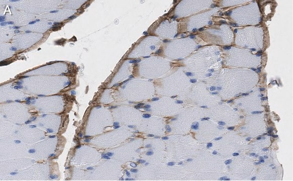
Immunohistochemistry Analysis: A representative lot immunostained fibroblastic cells in paraffin-embedded gastrocnemius section from a patient with myopathic condition (Courtesy of Duy Minh Ha, University of Nebraska Medical Center, Omaha, NE)
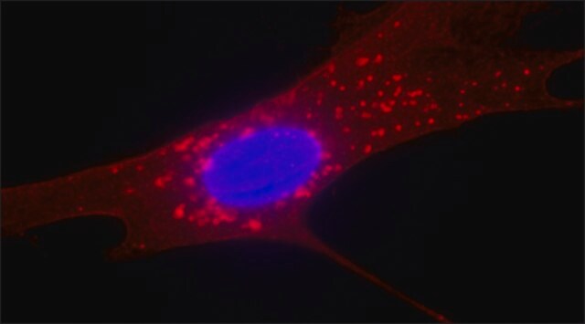
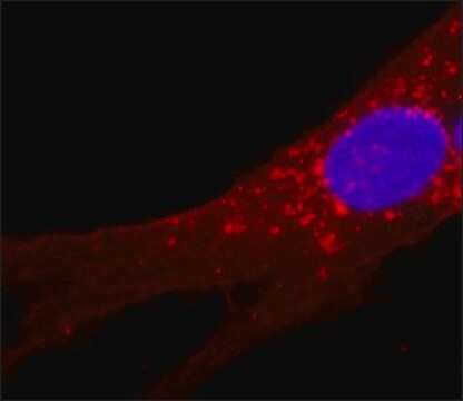
Immunocytochemistry Analysis: A representative lot immunostained human muscle fibroblasts by fluorescent immunocytochemistry (Agley, C.C., et al. (2015). J. Vis. Exp. (95):52049).
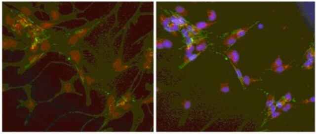
Immunocytochemistry Analysis: A representative lot immunostained MRC-5 fetal fibroblasts and normal human dermal fibroblasts (NHDF), but not fibrocytes or macrophages by immunocytochemistry (Bertolotto, C., et al. (2009). PLoS One. 4(10):e7475).
Flow Cytometry Analysis: A representative lot detected fibroblasts differentiated from human ESCs and iPSCs (Zhang, J., et al. (2012). Circ. Res. 111(9):1125-1136).
Immunofluorescence Analysis: A representative lot immunostained fibroblasts in human skeletal muscle cryosections by fluorescent immunohistochemistry (Agley, C.C., et al. (2013). J. Cell. Sci. 126(Pt 24):5610-5625).
Immunofluorescence Analysis: A representative lot immunostained the fibrous stroma and vessels in acetone-fixed, frozen human tissue sections by fluorescent immunohistochemistry (Haynes, B.F., et al. (1984). J. Exp. Med. 159(4):1149-1168).
Research Category
Inflammation & Immunology
Research Sub Category
Immunoglobulins & Immunology
This Anti-Fibroblasts Antibody, clone TE is validated for use in Flow Cytometry (FC), Immunocytochemistry (ICC), Immunofluorescence (IF) and Immunohistochemistry (IHC) for the detection of Fibroblasts.

Physical form

Format: Purified
The monoclonal is presented at a concentration of 100 μg/mL in phosphate buffered saline containing 10 mM sodium azide and 1 mg/mL bovine serum albumin. We recommend that each laboratory determine an optimum working titre for use in its particular application.

Storage and Stability

For use within 1 month of purchase store at +4°C, for long term storage aliquot antibody into small volumes and store at -20°C.

Legal Information

CHEMICON is a registered trademark of Merck KGaA, Darmstadt, Germany

Disclaimer

Unless otherwise stated in our catalog or other company documentation accompanying the product(s), our products are intended for research use only and are not to be used for any other purpose, which includes but is not limited to, unauthorized commercial uses, in vitro diagnostic uses, ex vivo or in vivo therapeutic uses or any type of consumption or application to humans or animals.

Not finding the right product?  

Try our Product Selector Tool.

Storage Class Code

12 - Non Combustible Liquids

WGK

WGK 2

Flash Point(F)

Not applicable

Flash Point(C)

Not applicable


Regulatory Listings

Regulatory Listings are mainly provided for chemical products. Only limited information can be provided here for non-chemical products. No entry means none of the components are listed. It is the user’s obligation to ensure the safe and legal use of the product.

JAN Code

CBL271:


Certificates of Analysis (COA)

Search for Certificates of Analysis (COA) by entering the products Lot/Batch Number. Lot and Batch Numbers can be found on a product’s label following the words ‘Lot’ or ‘Batch’.

Already Own This Product?

Find documentation for the products that you have recently purchased in the Document Library.

Visit the Document Library

The human thymic microenvironment.
Haynes, B F
Advances in Immunology, 36, 87-142 (1984)
Intrathymic and extrathymic T cell maturation.
Stutman, O
Immunological Reviews, 42, 138-184 (1978)
Katharine M Hinchcliff et al.
Journal of plastic, reconstructive & aesthetic surgery : JPRAS, 70(5), 568-576 (2017-03-28)
AlloDerm RTU® and AlloMaxTM are two acellular dermal matrices (ADMs) used in implant-based breast reconstruction. In this study, we examined whether different processing methods for the ADMs lead to a disparity in histologic, clinical, and financial outcomes after breast reconstruction.
Yusuke Hirano et al.
International journal of urology : official journal of the Japanese Urological Association, 30(1), 107-112 (2022-09-21)
Myofibroblast-dominant proliferation (relative to fibroblast proliferation) is the key process in urethral fibrosis, but its association with clinical features is not understood. We conducted a histological analysis of urethral strictures and examined the association between myofibroblast proliferation and stricture characteristics.
Duy M Ha et al.
Journal of translational medicine, 14, 39-39 (2016-02-06)
Lower leg ischemia, myopathy, and limb dysfunction are distinguishing features of peripheral artery disease (PAD). The myopathy of PAD is characterized by myofiber degeneration in association with extracellular matrix expansion, and increased expression of transforming growth factor-beta 1 (TGF-β1; a

Our team of scientists has experience in all areas of research including Life Science, Material Science, Chemical Synthesis, Chromatography, Analytical and many others.

Contact Technical Service