Passa al contenuto
Merck
Tutte le immagini(1)

Documenti fondamentali

247M-9

Sigma-Aldrich

EMA (E29) Mouse Monoclonal Antibody

Autenticatiper visualizzare i prezzi riservati alla tua organizzazione & contrattuali


About This Item

Codice UNSPSC:
12352203
NACRES:
NA.41

Origine biologica

mouse

Livello qualitativo

100
500

Coniugato

unconjugated

Forma dell’anticorpo

culture supernatant

Tipo di anticorpo

primary antibodies

Clone

E29, monoclonal

Descrizione

For In Vitro Diagnostic Use in Select Regions (See Chart)

Stato

buffered aqueous solution

Reattività contro le specie

human

Confezionamento

vial of 0.1 mL concentrate (247M-94)
vial of 0.5 mL concentrate (247M-95)
bottle of 1.0 mL predilute (247M-97)
vial of 1.0 mL concentrate (247M-96)
bottle of 7.0 mL predilute (247M-98)

Produttore/marchio commerciale

Cell Marque

tecniche

immunohistochemistry (formalin-fixed, paraffin-embedded sections): 1:100-1:500

Isotipo

IgG2aκ

Controllo

breast

Condizioni di spedizione

wet ice

Temperatura di conservazione

2-8°C

Visualizzazione

cytoplasmic, membranous

Informazioni sul gene

human ... MUC1(4582)

Categorie correlate

Descrizione generale

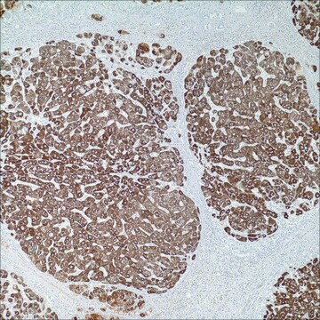
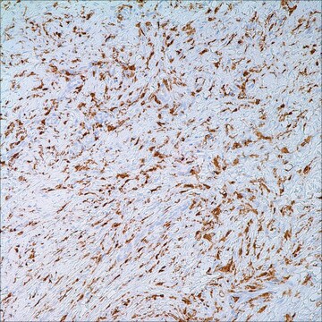
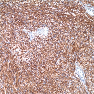
Anti-EMA antibody is a useful marker for staining many carcinomas. It stains normal and neoplastic cells from various tissues, including mammary epithelium, sweat glands and squamous epithelium. Hepatocellular carcinoma, adrenal carcinoma and embryonal carcinomas are consistently EMA negative, so keratin positivity with negative EMA favors one of these tumors. EMA is frequently positive in meningioma, which can be useful when distinguishing it from other intracranial neoplasms, e.g. Schwannomas. The absence of EMA can also be of value since negative EMA staining is characteristic of some tumors including adrenal carcinoma, seminomas, paraganglioma and hepatoma.

Qualità


IVD

IVD

IVD

RUO

Linkage

EMA Positive Control Slides, Product No. 247S, are available for immunohistochemistry (formalin-fixed, paraffin-embedded sections).

Stato fisico

Solution in Tris Buffer, pH 7.3-7.7, with 1% BSA and <0.1% Sodium Azide

Nota sulla preparazione

Download the IFU specific to your product lot and formatNote: This requires a keycode which can be found on your packaging or product label.

Altre note

For Technical Service please contact: 800-665-7284 or email: service@cellmarque.com

Note legali

Cell Marque is a trademark of Merck KGaA, Darmstadt, Germany

Non trovi il prodotto giusto?  

Prova il nostro Motore di ricerca dei prodotti.


Scegli una delle versioni più recenti:

Certificati d'analisi (COA)

Lot/Batch Number

Non trovi la versione di tuo interesse?

Se hai bisogno di una versione specifica, puoi cercare il certificato tramite il numero di lotto.

Possiedi già questo prodotto?

I documenti relativi ai prodotti acquistati recentemente sono disponibili nell’Archivio dei documenti.

Visita l’Archivio dei documenti

M Fraga et al.
American journal of clinical pathology, 103(1), 82-89 (1995-01-01)
Bone marrow involvement by anaplastic large cell anaplastic large cell (ALC) lymphoma can be difficult to detect on routine morphologic examination alone. In a series of 42 patients with ALC lymphoma, the authors analyzed: (1) the usefulness of a limited
G S Pinkus et al.
American journal of clinical pathology, 85(3), 269-277 (1986-03-01)
Epithelial membrane antigen and keratin proteins represent markers of epithelial differentiation that may be detected in routine formalin-fixed, paraffin-embedded tissues. Eighty-seven neoplasms, including 48 adenocarcinomas of various types, squamous and transitional cell carcinomas, small-cell anaplastic carcinomas, carcinoid tumors, mesotheliomas, hepatomas
R L Attanoos et al.
Histopathology, 43(3), 231-238 (2003-08-28)
To evaluate the expression of the intermediate filament desmin in reactive mesothelium and malignant mesothelioma and to compare its utility with five other previously reported immunomarkers claimed to be of use in distinguishing reactive from neoplastic mesothelium. Sixty cases of
D P Dearnaley et al.
British journal of cancer, 44(1), 85-90 (1981-07-01)
We have developed a technique for the immunocytochemical staining of marrow smears using antiserum to epithelial membrane antigen (EMA). This membrane component is confined to, but widely distributed in, epithelial tissues and tumours derived from them, and is strongly expressed
J S Lee et al.
Acta cytologica, 40(4), 631-636 (1996-07-01)
This study was designed to assess whether a new panel of antibodies is a useful adjunct in the differential diagnosis of carcinoma and reactive mesothelial cells. Complete, one-hour immunohistochemistry using antibodies against cytokeratin (CK), carcinoembryonic antigen (CEA), epithelial membrane antigen

Il team dei nostri ricercatori vanta grande esperienza in tutte le aree della ricerca quali Life Science, scienza dei materiali, sintesi chimica, cromatografia, discipline analitiche, ecc..

Contatta l'Assistenza Tecnica.