Passa al contenuto
Merck
Tutte le immagini(4)

Documenti fondamentali

MABT1340

Sigma-Aldrich

Anti-Lamin A/C Antibody, clone 2A1

clone 2A1, from mouse

Sinonimo/i:

70 kDa Lamin, Renal carcinoma antigen NY-REN-32

Autenticatiper visualizzare i prezzi riservati alla tua organizzazione & contrattuali


About This Item

Codice UNSPSC:
12352203
eCl@ss:
32160702
NACRES:
NA.41

Origine biologica

mouse

Forma dell’anticorpo

purified immunoglobulin

Tipo di anticorpo

primary antibodies

Clone

2A1, monoclonal

Reattività contro le specie

mouse, monkey, hamster, human

Confezionamento

antibody small pack of 25 μg

tecniche

immunocytochemistry: suitable
immunofluorescence: suitable
immunoprecipitation (IP): suitable
western blot: suitable

Isotipo

IgG2bκ

N° accesso NCBI

N° accesso UniProt

modifica post-traduzionali bersaglio

unmodified

Informazioni sul gene

human ... LMNA(4000)

Descrizione generale

Prelamin-A/C (UniProt: P02545) is encoded by the LMNA (also known as LMN1) gene (Gene ID: 4000) in human. It is cleaved into Lamin-A/C (also known as 70 kDa Lamin, Renal carcinoma antigen NY-REN-32). Lamins are components of the nuclear lamina that provide a framework for the nuclear envelope and may also interact with chromatin. Lamin A and C are present in equal amounts in the lamina of mammals. Plays an important role in nuclear assembly, chromatin organization, nuclear membrane and telomere dynamics. Lamin A is initially synthesized as prelamin A that undergoes several modifications in the carboxyl terminal region that allow incorporation of prelamin A into the nuclear envelope and its subsequent processing into the mature lamin A. Cleavage of 15 residues (aa 647-662) by ZMPSTE24/FACE1 generates the final protein product. Unlike mature lamin A, prelamin A accumulates as discrete and localized foci at the nuclear periphery. Prelamin-A/C can accelerate smooth muscle cell senescence. It can act to disrupt mitosis and induce DNA damage in vascular smooth muscle cells (VSMCs), leading to mitotic failure, genomic instability, and premature senescence. Mutations in LMNA gene are known to cause Emery-Dreifuss muscular dystrophy that is characterized by weakness and atrophy of muscle without involvement of the nervous system. Some mutations have also been linked to familial type of lipodystrophy characterized by the loss of subcutaneous adipose tissue in the lower parts of the body. (Ref.: Casasola, A., et al. (2016). Nucleus 7(1); 84-102).

Specificità

Clone 2A1 detects the Lamin A/C species in the nuclear interior in multiple species. It targets the Rod domain of Lamin A/C.

Immunogeno

Purified protein corresponding to the rod domain for human Lamin A/C.

Applicazioni

Anti-Lamin A/C, clone 2A1, Cat. No. MABT1340, is a highly specific mouse monoclonal antibody that targets Lamin A/C and has been tested for use in Immunocytochemistry, Immunofluorescence, Immunoprecipitation, and Western Blotting.
Immunoprecipitation Analysis: A representative immunoprecipitated Lamin A/C in culture supernatant (Data courtesy of Marie Lang, M.D., Stefan Schuchner, Ph.D. and Egon Ogris, M.D., Medical University of Vienna, Austria).


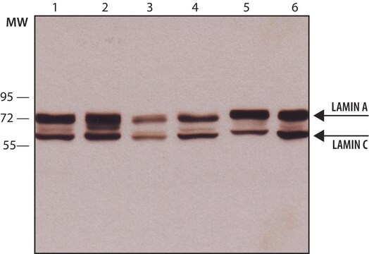
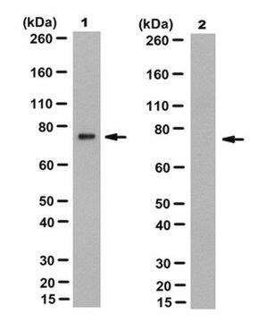
Western Blotting Analysis: A representative lot detected Lamin A/C in culture supernatants of various cell lines (Data courtesy of Marie Lang, M.D., Stefan Schuchner, Ph.D. and Egon Ogris, M.D., Medical University of Vienna, Austria).


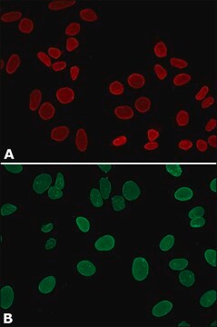
Immunocytochemistry Analysis: A 1:1,000 dilution from a representative lot detected Lamin A/C in HeLa cells.


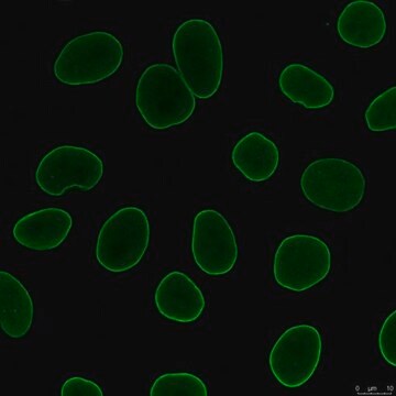
Immunofluorescence Analysis: A representative lot detected Lamin A/C in the nuclear interior of HeLa cells (Data courtesy of Marie Lang, M.D., Stefan Schuchner, Ph.D. and Egon Ogris, M.D., Medical University of Vienna, Austria).
Research Category
Cell Structure

Qualità

Evaluated by Western Blotting in HeLa cell lysate.

Western Blotting Analysis: 0.2 µg/mL of this antibody detected Lamin A/C in HeLa cell lysate.

Descrizione del bersaglio

74 and 65 kDa observed. 74.14 kDa and 65.14 kDa for Lamin A and C. Uncharacterized bands may be observed in some lysate(s).

Stato fisico

Format: Purified
Protein G purified
Purified mouse monoclonal antibody IgG2b in buffer containing 0.1 M Tris-Glycine (pH 7.4), 150 mM NaCl with 0.05% sodium azide.

Stoccaggio e stabilità

Stable for 1 year at 2-8°C from date of receipt.

Altre note

Concentration: Please refer to lot specific datasheet.

Esclusione di responsabilità

Unless otherwise stated in our catalog or other company documentation accompanying the product(s), our products are intended for research use only and are not to be used for any other purpose, which includes but is not limited to, unauthorized commercial uses, in vitro diagnostic uses, ex vivo or in vivo therapeutic uses or any type of consumption or application to humans or animals.

Non trovi il prodotto giusto?  

Prova il nostro Motore di ricerca dei prodotti.


Certificati d'analisi (COA)

Cerca il Certificati d'analisi (COA) digitando il numero di lotto/batch corrispondente. I numeri di lotto o di batch sono stampati sull'etichetta dei prodotti dopo la parola ‘Lotto’ o ‘Batch’.

Possiedi già questo prodotto?

I documenti relativi ai prodotti acquistati recentemente sono disponibili nell’Archivio dei documenti.

Visita l’Archivio dei documenti

Il team dei nostri ricercatori vanta grande esperienza in tutte le aree della ricerca quali Life Science, scienza dei materiali, sintesi chimica, cromatografia, discipline analitiche, ecc..

Contatta l'Assistenza Tecnica.