Passa al contenuto
Merck
Tutte le immagini(3)

Documenti fondamentali

MABE175

Sigma-Aldrich

Anti-PML Isoform II Antibody, clone 1A8.1

clone 1A8.1, from mouse

Sinonimo/i:

PML-2, PML-II, Protein PML isoform II, Promyelocytic leukemia protein isoform II, RING finger protein 71 isoform II, TRIM19kappa, Tripartite motif-containing protein 19 isoform II

Autenticatiper visualizzare i prezzi riservati alla tua organizzazione & contrattuali


About This Item

Codice UNSPSC:
12352203
eCl@ss:
32160702
NACRES:
NA.43

Origine biologica

mouse

Livello qualitativo

Forma dell’anticorpo

purified immunoglobulin

Tipo di anticorpo

primary antibodies

Clone

1A8.1, monoclonal

Reattività contro le specie

human

tecniche

immunocytochemistry: suitable
western blot: suitable

Isotipo

IgG1κ

N° accesso NCBI

N° accesso UniProt

modifica post-traduzionali bersaglio

unmodified

Informazioni sul gene

human ... PML(5371)

Descrizione generale

Protein PML (UniProt P29590; also known as Promyelocytic leukemia protein, RING finger protein 71, Tripartite motif-containing protein 19) is encoded by the PML (also known as MYL, PP8675, RNF71, TRIM19) gene (Gene ID 5371) in human. The human PML gene/protein adopted its name when it was originally discovered as an RARα fusion protein caused by chromosomal translocation in acute promyelocytic leukemia (APL) patients. In normal cells, the PML protein interacts with multiple protein partners in the nucleus to form PML nuclear bodies (PML-NBs) that mediate a variety of cellular functions including senescence, apoptosis, protein degradation, telemere stasbility, and antiviral defense. At least 12 PML isoforms are generated by alternative splicings, including six nuclear isoforms (PML-I to PML-VI) and the cytoplasmic isoform PML-VII (a.k.a. PML-VIIB), other spliced isoforms without exon 6-coding sequence (a.a. 467-552) are likewise expected to be cytoplasmic due to the abscence of nuclear localization signal (NLS) encoded by exon 6. All isoforms share the same N-terminal region (a.a. 1-419) containing the RBCC/tripartite motif and may have related functions due to their common functional RBCC/TRIM domain, while increasing evidences also suggest that each PML isoform possesses distinct functions mediated by its specific C-terminal sequence. Isoform PML-1 (a.k.a. PML-I, TRIM19alpha; UniProt P29590-1) and PML-2 (a.k.a. PML-II or TRIM19kappa; UniProt P29590-8) share the same N-terminal 570-a.a. sequence (encoded by exons 1 to 7a), however PML-1 derives its remaining C-terminal sequence (a.a. 571-882) from exons 8a & 9, while PML-II derives its (a.a. 571-829) from exon 7b with a different reading frame as that of PML-V.

Specificità

Clone 1A8.1 was raised against an C-terminal region sequence unique to PML isoform II (PML-2), but not present in the other 11 PML spliced isoforms reported by UniProt (P29590).

Immunogeno

Epitope: C-terminal region.
KLH-conjugated linear peptide corresponding to a C-terminal sequence of human PML Isoform II.

Applicazioni

Anti-PML Isoform II Antibody, clone 1A8.1 is an antibody against PML Isoform II for use in Western Blotting, Immunocytochemistry.
Immunocytochemistry Analysis: 40 µg/mL from a representative lot immunostained 10% neutral-buffered formalin-fixed HEK293 cells transfected with human PML isoform II by non-fluorescent immunocytochemistry (Courtesy of Professor Ygal Haupt, Peter MacCallum Cancer Centre, East Melbourne, Australia).
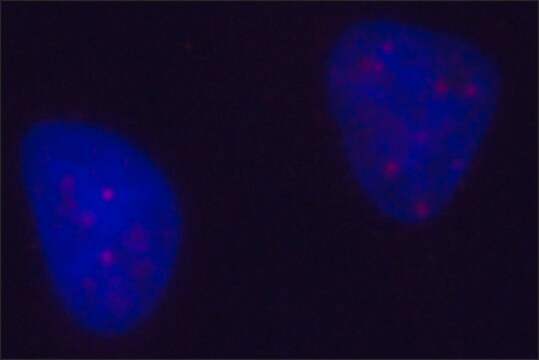
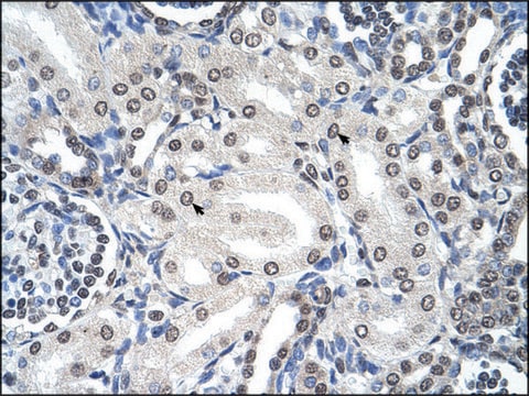
Immunocytochemistry Analysis: 10 µg/mL from a representative lot immunostained 4% paraformaldehyde-fixed HEK293 cells transfected with human PML isoform II by fluorescent immunocytochemistry (Courtesy of Professor Ygal Haupt, Peter MacCallum Cancer Centre, East Melbourne, Australia).
Research Category
Epigenetics & Nuclear Function
Research Sub Category
Chromatin Biology

Qualità

Evaluated by Western Blotting in lysate from PML-2 transfected HEK293 cells.

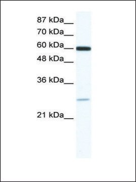
Western Blotting Analysis: 0.5 µg/mL of this antibody detected the exogenously expressed human PML-2 in 10 µg of lysate from transfected HEK293 cells.

Descrizione del bersaglio

~110 kDa observed. 90.72 kDa calculated. Uncharacterized band(s) may appear in some lysates.

Stato fisico

Format: Purified
Protein G Purified
Purified mouse monoclonal IgG1κ antibody in buffer containing 0.1 M Tris-Glycine (pH 7.4), 150 mM NaCl with 0.05% sodium azide.

Stoccaggio e stabilità

Stable for 1 year at 2-8°C from date of receipt.

Altre note

Concentration: Please refer to lot specific datasheet.

Esclusione di responsabilità

Unless otherwise stated in our catalog or other company documentation accompanying the product(s), our products are intended for research use only and are not to be used for any other purpose, which includes but is not limited to, unauthorized commercial uses, in vitro diagnostic uses, ex vivo or in vivo therapeutic uses or any type of consumption or application to humans or animals.

Non trovi il prodotto giusto?  

Prova il nostro Motore di ricerca dei prodotti.

Codice della classe di stoccaggio

12 - Non Combustible Liquids

Classe di pericolosità dell'acqua (WGK)

WGK 1

Punto d’infiammabilità (°F)

Not applicable

Punto d’infiammabilità (°C)

Not applicable


Certificati d'analisi (COA)

Cerca il Certificati d'analisi (COA) digitando il numero di lotto/batch corrispondente. I numeri di lotto o di batch sono stampati sull'etichetta dei prodotti dopo la parola ‘Lotto’ o ‘Batch’.

Possiedi già questo prodotto?

I documenti relativi ai prodotti acquistati recentemente sono disponibili nell’Archivio dei documenti.

Visita l’Archivio dei documenti

Il team dei nostri ricercatori vanta grande esperienza in tutte le aree della ricerca quali Life Science, scienza dei materiali, sintesi chimica, cromatografia, discipline analitiche, ecc..

Contatta l'Assistenza Tecnica.